Nodular amyloidosis
Nodular amyloidosis
Clinical features In this rare variant, which is more common in females, pink–brown single or multiple nodules develop on the trunk, extremities, genitalia, face or scalp (Fig. 13.61).1–13 Bilateral nodular amyloidosis of the eyelids in the absence
The deposits of amyloid are present in both the papillary and reticular dermis and may involve the subcutaneous fat (Figs 13.62–13.64). Sometimes
A
B
582 Degenerative and metabolic diseases
the vasculature and nerve sheaths are affected (Figs 13.65–13.67).2 Characteristically, plasma cells are seen around blood vessels and at the margin of the amyloid deposits (Fig. 13.68).4,38 Rarely, an associated foreign body giant cell reaction with phagocytosis of amyloid and/or calcification are evident.4,6
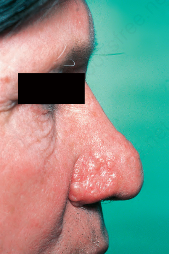
Fig. 13.61 Nodular amyloidosis: an irregular infiltrated plaque limited to the nose.
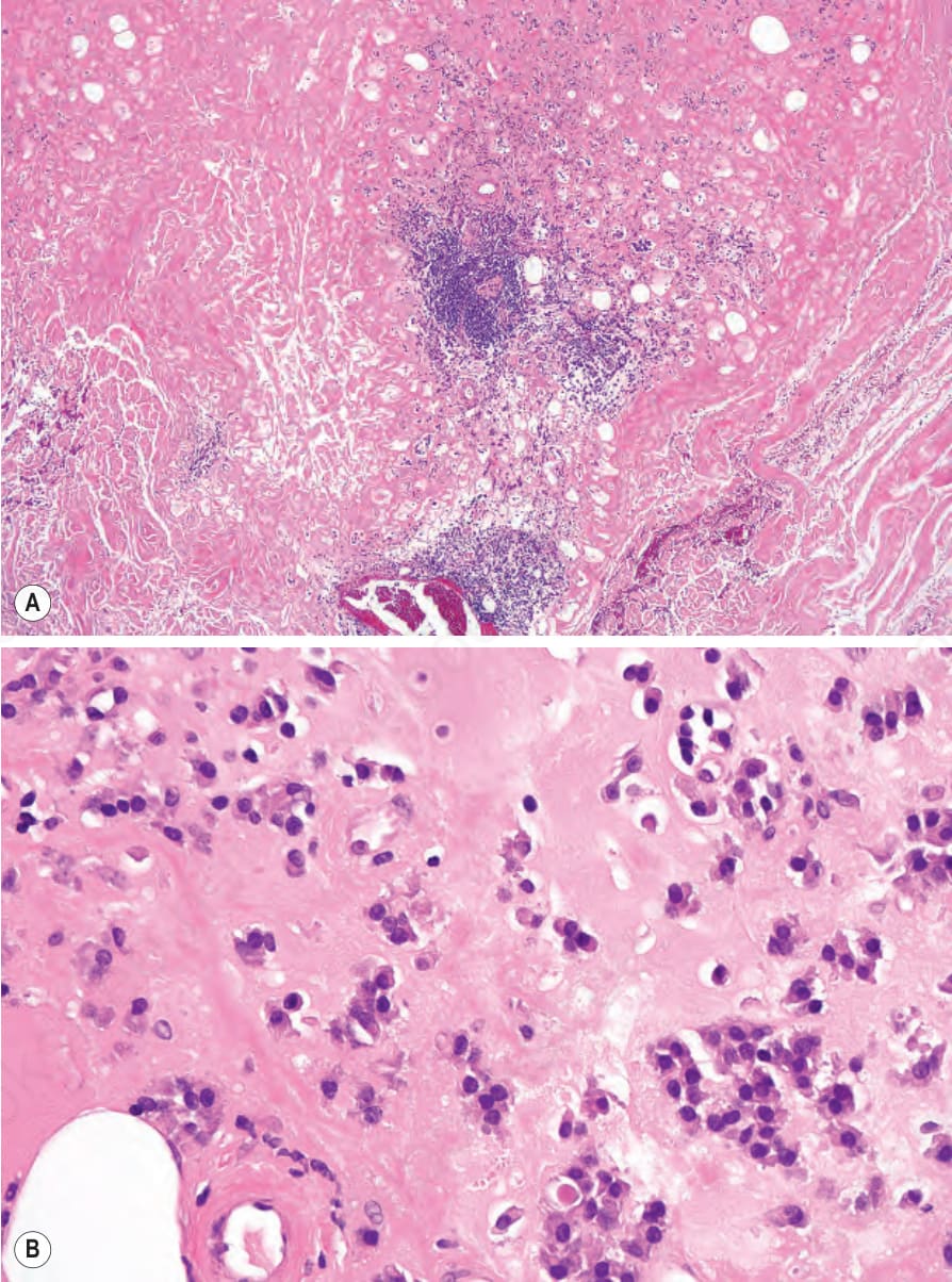
Fig. 13.62 Nodular amyloidosis: (A) massive deposits of amyloid are present in the dermis; (B) there is a heavy associated plasma cell infiltrate.
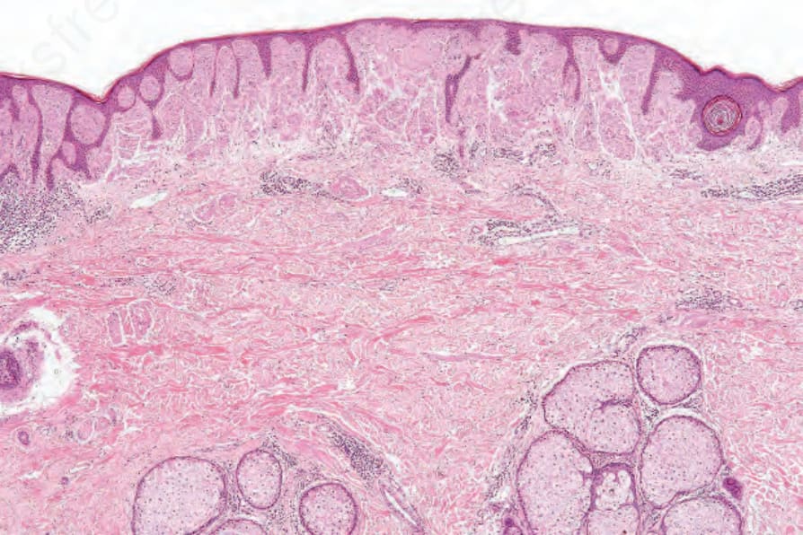
Fig. 13.63 Nodular amyloidosis: in this example there is a broad bandlike deposit in the upper dermis.
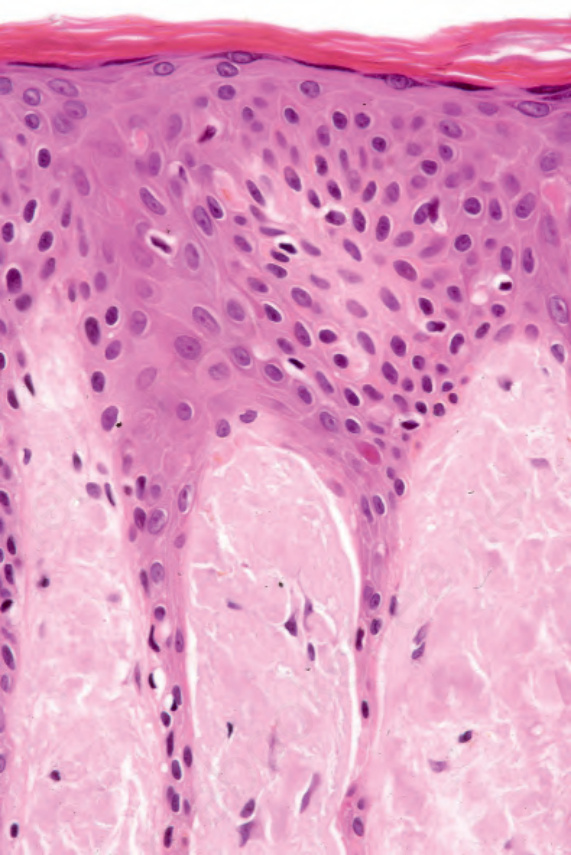
Fig. 13.64 Nodular amyloid: the amyloid deposits fill the papillary dermis.
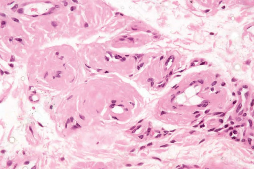
Fig. 13.65 Nodular amyloidosis: amyloid deposits have thickened the blood vessel walls.
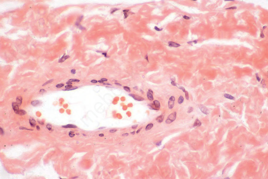
Fig. 13.66 Nodular amyloid: the deposits are strongly Congo red positive.
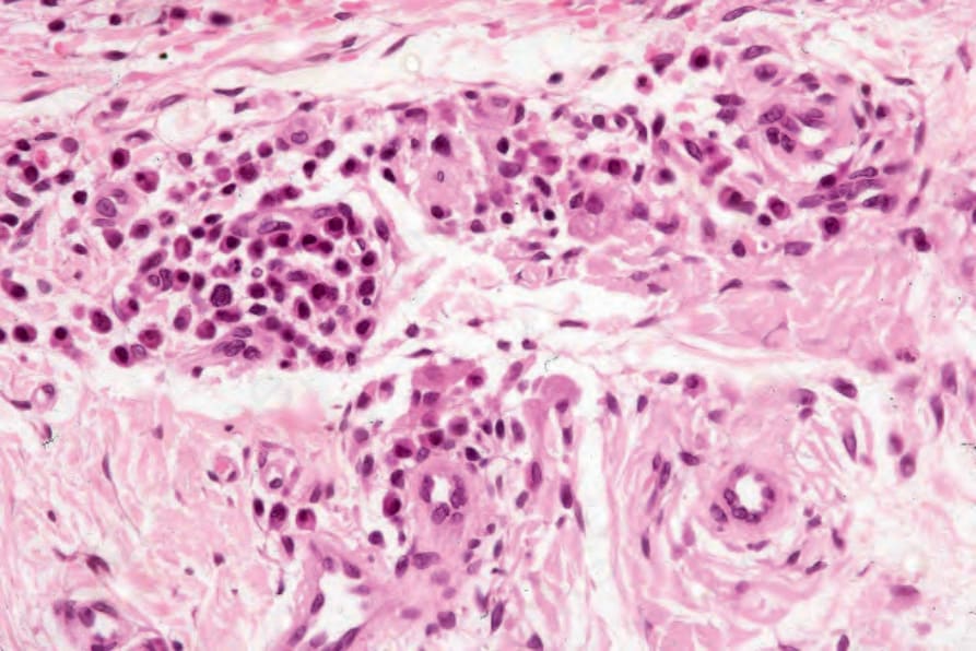
Fig. 13.68 Nodular amyloidosis: there is a conspicuous plasma cell infiltrate.