The amyloidoses
The amyloidoses
Amyloidosis is characterized by the extracellular deposition of a protein associated with particular tinctorial and ultrastructural properties. The amyloidoses are classified according to whether the amyloid deposition is systemic or localized (Table 13.3).
The most characteristic staining patterns of amyloid are seen with Congo red or Dylon (cotton dye pagoda red No. 9), which show apple-green birefringence under polarized light (Fig. 13.36).1 Unfortunately, this is not specific, and green birefringence may also be seen with collagen and in colloid milium, porphyria, and lipoid proteinosis. Amyloid deposits, which are PAS positive, may also be identified by the cotton dye Sirius red, or metachromatically using methyl or cresyl violet.2 Further confirmatory evidence can be obtained by staining with thioflavine-T and examination using fluorescence microscopy or by immunocytochemistry (see below) (Fig. 13.37).
B
Amyloid shows characteristic and specific electron microscopic features of rigid, straight, nonbranching amyloid filaments with a diameter of 6–10 nm showing a hollow core on cross-section (Fig. 13.38).3 They are haphazardly distributed, lack the cross-banding of collagen, and are embedded in an electron-dense amorphous ground substance, which is probably composed of polysaccharides.
X-ray diffraction and infrared spectroscopy reveal a beta-pleated antiparallel configuration.4,5 Fibrils with a beta-pleated configuration are insoluble and highly resistant to proteolysis. This, combined with a lack of immunogenicity, results in their persistence at the site of deposition and subsequent tissue-damaging effects.
All forms of amyloid contain up to 14% by dry weight of a nonfibrillary protein, the serum amyloid P (SAP) component.2,6 The function of SAP is unknown, but it has been suggested that it may be primarily involved in the deposition and maintenance of the fibrillary components.2 Its presence, identified immunohistochemically, is a useful adjunct to the diagnosis of amyloidosis.7 However, it should be appreciated that the antibody also labels degenerate elastic fibers. The fibrillary component, however, may be derived in very different ways in each of the recognized types of amyloidosis:8
• In primary and myeloma-associated amyloidoses it consists of immunoglobulin light chains (most often of lambda type, or a part thereof).
• In the secondary form the fibrillary component is composed of amyloid A protein, which is derived from a normal serum constituent known as serum amyloid A protein. This serum protein, which is an HDL3-associated apolipoprotein, is an acute phase reactant.2,8
573 The amyloidoses
A
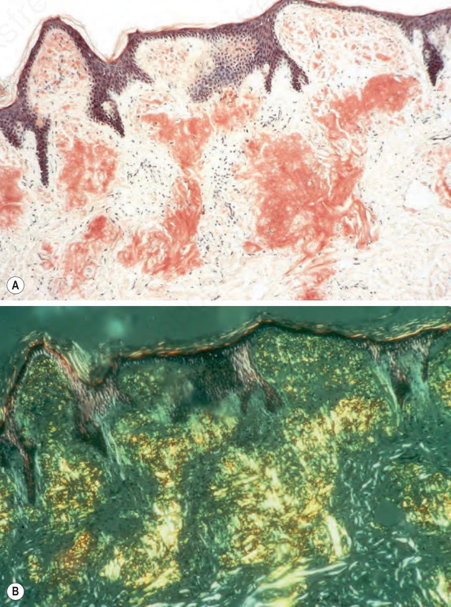
Fig. 13.36 Cutaneous amyloidosis: (A) positive staining with Congo red; (B) there is intense apple-green birefringence when viewed with polarized light.
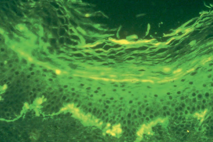
Fig. 13.37 Cutaneous amyloidosis: positive immunofluorescence just beneath the epidermis in a case of macular amyloid (thioflavine-T).
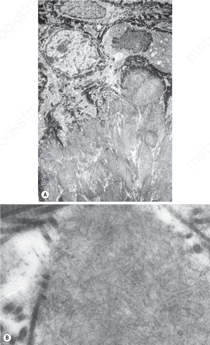
Fig. 13.38 Cutaneous amyloidosis: (A) electron micrograph of macular amyloidosis showing nodular deposits in the superficial dermis; (B) the characteristic randomly orientated, straight, nonbranching appearance of amyloid filaments.
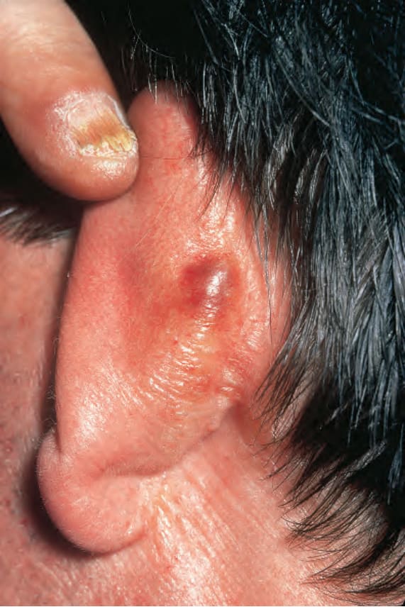
Fig. 13.39 Primary systemic amyloidosis: a waxy nodule is present behind the ear. Note the purpura. By courtesy of R.A. Marsden, MD, St George’s Hospital, London, UK.
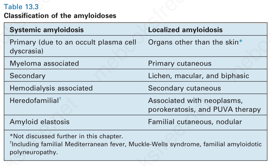
Table 13.3 Classification of the amyloidoses