Tendinous xanthomata
Tendinous xanthomata
Clinical features Tendinous xanthomata, which are associated with raised LDL levels, are slowly enlarging subcutaneous tumors that occur in tendons (especially those of the hands, knees, elbows, and the Achilles tendon), ligaments, fascia, and periosteum (Figs 13.8 and 13.9).1 The overlying skin, which appears normal, is freely moveable over the surface and small tendon xanthomata may be difficult to palpate.1 The lesions characteristically ‘move with the tendons’ and are thought to be trauma related.2 The presence of these xanthomata is most frequently a feature of heterozygous familial (LDL receptor deficiency) hypercholesterolemia.2–4 There is a high risk of
564 Degenerative and metabolic diseases
associated coronary atherosclerosis. A meta-analysis demonstrated a threefold increased risk of cardiovascular disease in patients with familial hypercholesterolemia and tendinous xanthomata compared to those without cutaneous lesions.5 Tendinous xanthomata are also seen in familial combined hyperlipidemia, normocholesterolemic states such as cerebrotendinous xanthomatosis (cholestanolosis) and β-sitosterolemia, and the nephrotic syndrome.2,6–10 Cerebrotendinous xanthomatosis is an autosomal recessive disease caused by a mutation in CYP27A1, the sterol 27-hydroxylase gene that is an important regulator of brain cholesterol homoestasis.11 This results in xanthoma in the brain as well as tendons.
Clinically, the lesions, which may be mistaken for gouty tophi and rheumatoid nodules, are sometimes found in association with tuberous xanthomata and xanthelasmata.
Histologic features Tendinous xanthomata are composed of multiple nodules containing xanthoma cells, accompanied in early lesions by an admixture of inflammatory cells including non-lipidized histiocytes, lymphocytes, and neutrophil polymorphs. The deposits in tendinous xanthoma are doubly refractile to polarized light (Fig. 13.10). Older lesions are characteristically associated with fibrosis.
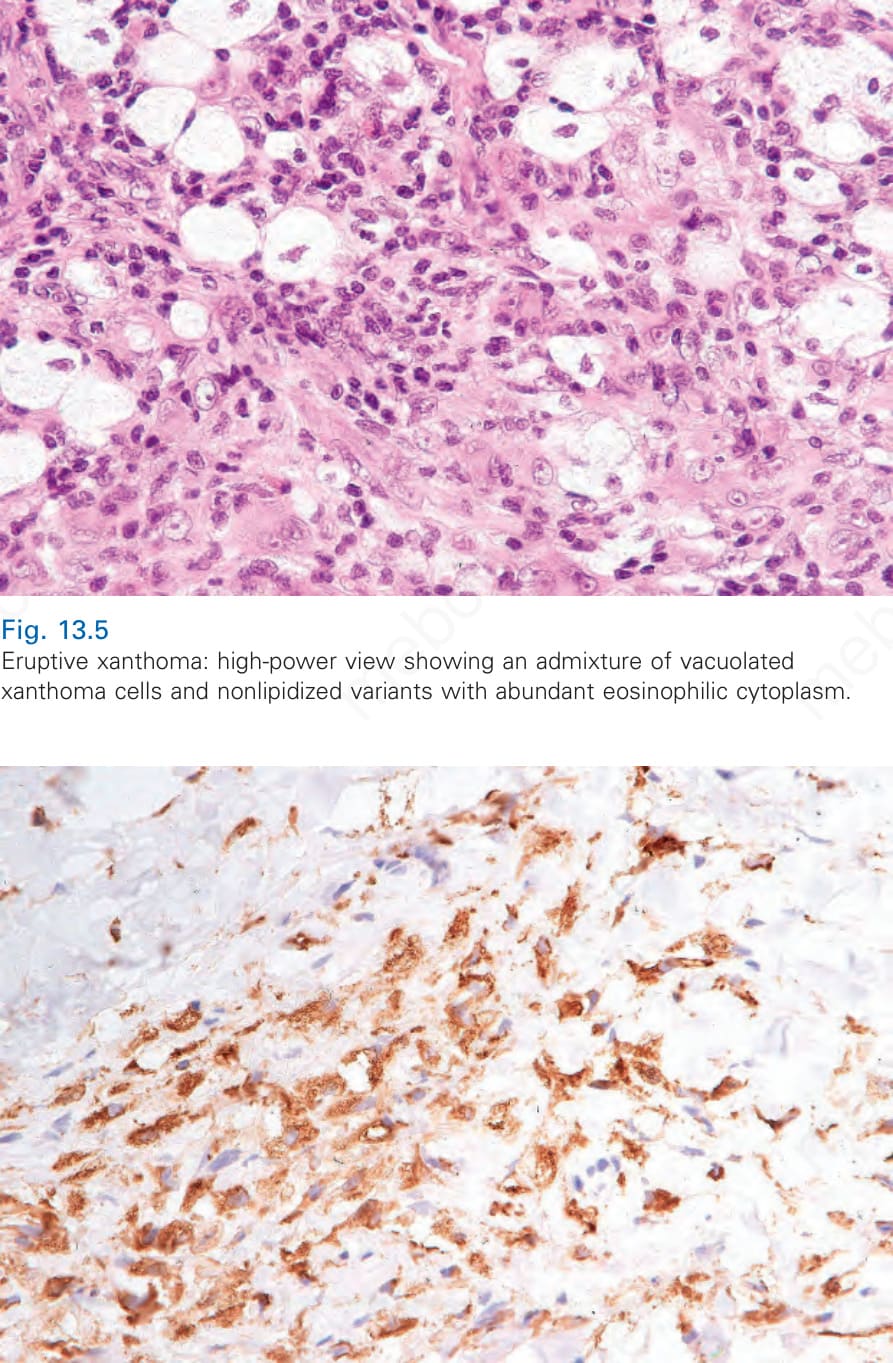
Fig. 13.6 Eruptive xanthoma: the histiocytes express CD68.
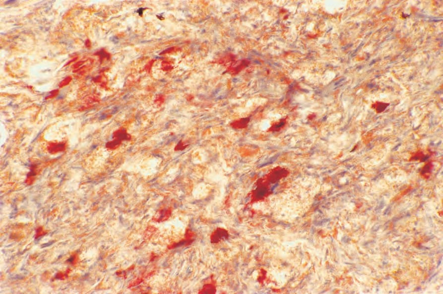
Fig. 13.7 Eruptive xanthoma: the lipid within the macrophages stains positively with oil red O.
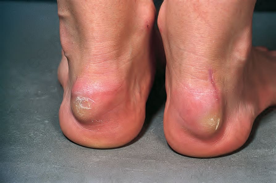
Fig. 13.8 Tendinous xanthoma: typical nodules on the heels. These lesions are often related to trauma; the Achilles tendon is a classical site. By courtesy of A.F. Lant, MD, and J. Dequeker, MD, London, UK.

Fig. 13.9 Tendinous xanthoma: xanthomata are present overlying the knuckles. By courtesy of the Institute of Dermatology, London, UK.
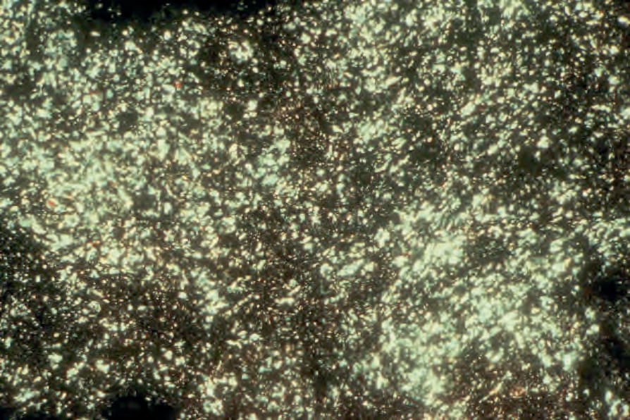
Fig. 13.10 Tendinous xanthoma: intense birefringence of deposits in polarized light (oil red O).
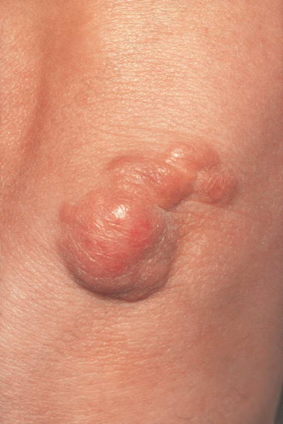
Fig. 13.11 Tuberous xanthoma: firm erythematous nodules over the elbow. By courtesy of R.A. Marsden, MD, St George’s Hospital, London, UK.