Cloacogenic carcinoma
Cloacogenic carcinoma
This rare tumor presents in middle-aged women as a superficial ulcerated adenocarcinoma composed of colonic-type glands arising in direct continuity with vulval surface epithelium (Figs 12.267–12.269).1–4 It is independent of the perivulval glands and, by definition, direct extension or metastasis from an underlying large intestinal or visceral adenocarcinoma has been excluded. The clinical features are not distinctive but tumors may present as lesions simulating Bartholin gland infection.5 In a single case, the neoplastic glands also contained Paneth cells.6 The origin of this tumor is not known, but it is thought most probably to arise from an area of gastrointestinal metaplasia or from heterotopic intestinal tissue (Fig. 12.270). Vulval cloacogenic carcinoma should not be confused with the similarly named tumor of the anal canal. Tumor cells are positive for CK7 and CK20 and negative for estrogen and progesterone receptors.7
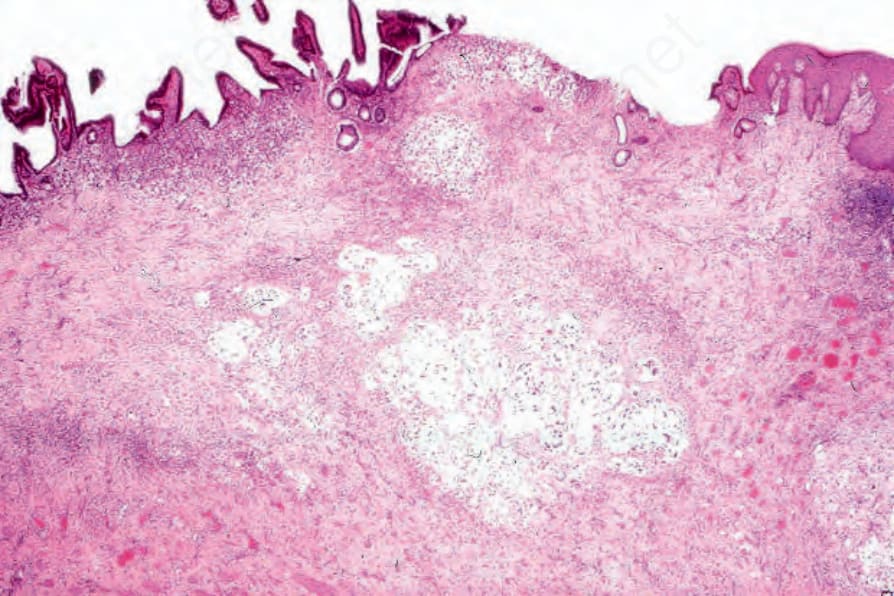
Fig. 12.267 Cloacogenic vulval adenocarcinoma: scanning view showing vulval squamous epithelium on the far right. Colonic epithelium is present on the left. Mucussecreting carcinoma extends throughout the underlying connective tissue.
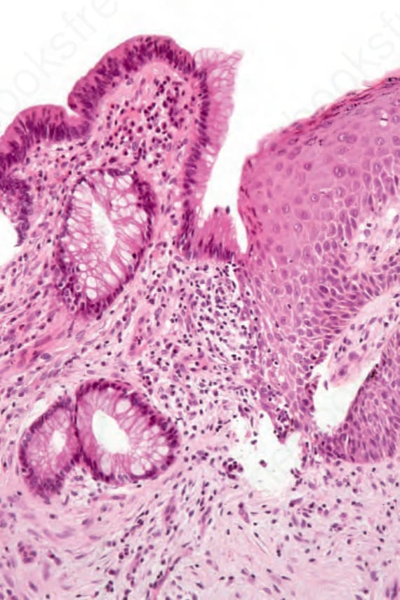
Fig. 12.270 Cloacogenic vulval adenocarcinoma: high-power view of the junction between squamous and colonic epithelium.