Lymphogranuloma venereum
Lymphogranuloma venereum
Clinical features Lymphogranuloma venereum is endemic in Asia, Africa, and South America and was considered to be rare in developed countries until several outbreaks were reported in a number of European countries and the United States, mainly in homosexuals and in the context of HIV infection.1,2 It is less common in women.3
The disease evolves in three stages:
• In stage 1 disease, the primary lesion develops 3–30 days after contact and is a small, transient, frequently asymptomatic papulovesicle or ulcer on the penis, scrotum, rectum, vulva (most commonly at the fourchette), vagina, and/or cervix (Figs 12.135 and 12.136).4 Primary lesions have been described on the fingers and tongue. Rarely, lymphogranuloma venereum has been reported presenting with a psoas abscess.5 Cat scratch disease may clinically simulate this disease.6
• Stage 2 develops within a few weeks of the primary lesion and consists of enlargement of the inguinal nodes. The pelvic lymph nodes may be enlarged in females. The lymphadenopathy is severe and initially painless and hard; later, the nodes (buboes) soften and discharge viscous pus. The tissue around the nodes becomes involved in the inflammatory process so that they become matted together. Along with lymphadenopathy, the patient may also experience malaise, joint pains, and hepatosplenomegaly. Erythema nodosum, light-sensitive eruptions, and erythema multiforme may complicate this phase and are more common in women.
• Stage 3 disease consists of complications of the early inflammatory changes. Involvement of the deep iliac and perirectal lymph nodes resulting from drainage from a high vaginal, posterior urethral, cervical,
The central necrosis is slowly absorbed and replaced by fibrosis. As a consequence, lymphedema develops distally. The lymphatics are typically inflamed and granulomata may be seen.
The diagnostic standard is now nucleic acid amplification testing.13 PCR may also be used to confirm the diagnosis.14
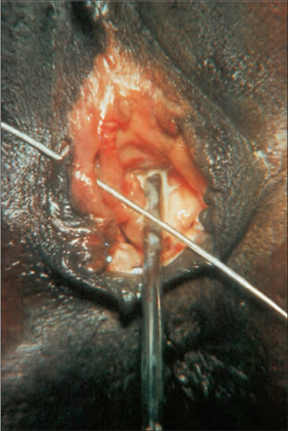
Fig. 12.135 Lymphogranuloma venereum: note the ulcer on the right labium majus. By courtesy of S. Lucas, MD, St. Thomas’ Hospital, London, UK.