Zoon balanitis
Zoon balanitis
Clinical features Zoon plasma cell balanitis (ZB) is a disorder of middle-aged and older uncircumcised males, although an analogous condition has been reported to afflict the vulva, mouth, lips, and epiglottis.1–7 Exceptionally, the disease may present in a circumcised male.5 True ZB is probably rare with many cases of LS being misdiagnosed as ZB. The presentation is classically indolent and asymptomatic. Well-demarcated, glistening, moist, bright red or brown patches involve the glans and visceral prepuce with sparing of the keratinized penile shaft and foreskin (Fig. 12.63). The navicular fossa may be involved. Other signs include dark red stippling – ‘cayenne pepper spots’ – and purpura with hemosiderin deposition, solitary or multiple lesions of differing sizes (guttate or nummular), characteristically symmetrical about the axis of the coronal sulcus, and ‘kissing’. Although vegetative and nodular presentations have been recorded, atypical or unusual morphology should be viewed with great suspicion and biopsied.8,9 The clinical differential diagnosis includes LS, erosive LP, psoriasis, seborrheic dermatitis, contact dermatitis, fixed drug eruption, secondary syphilis, histoplasmosis, erythroplasia of Queyrat, and Kaposi sarcoma.10,11 A confident clinical
Pathogenesis and histologic features Since Zoon’s original reports, there have been many accounts in the literature, but the etiology remains uncertain. The evidence suggests that ZB is a chronic, reactive, principally irritant dermatosis brought about by a dysfunctional prepuce. Retention of urine and squames and excessive frictional trauma (ZB is often located on the dorsal aspect of the glans and/or the adjacent prepuce, sites of maximal friction on foreskin retraction) create the irritation. There is no evidence of an infectious cause, and immunohistochemical findings suggest that ZB represents a non-specific polyclonal tissue reaction, consistent with an irritant dermatosis.6,7
Classically, the epidermis shows attenuation with absent granular and horny layers, and diamond- or lozenge-shaped basal cell keratinocytes with sparse dyskeratosis and spongiosis (Figs 12.64 and 12.65). In the dermis, there is a dense band of infiltration with plasma cells of variable density.13 Other signs include extravasated erythrocytes, hemosiderin, and vascular proliferation. Zoon stressed the presence of the plasma cell infiltrate in this condition, but in practice the plasma cell density can be very variable9,13,19
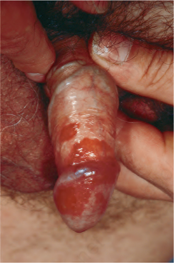
Fig. 12.63 Zoon balanitis: there is involvement of the glans, prepuce, and shaft of the penis. From Bunker C. Male Genital Skin Disease. Saunders Ltd./Elsevier 2004.
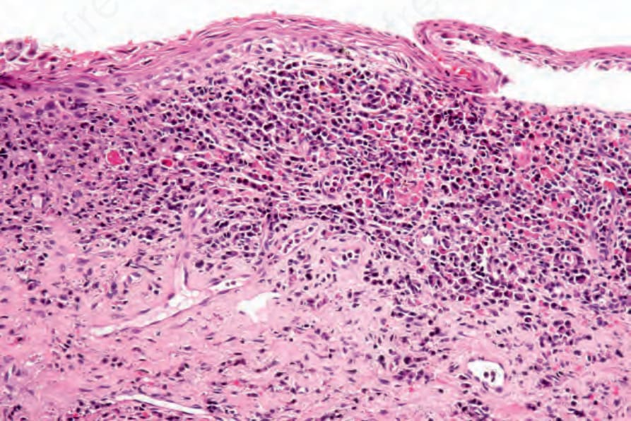
Fig. 12.64 Zoon balanitis: note the epidermal thinning, spongiosis, and a dense superficial inflammatory cell infiltrate.