Leiomyosarcoma
Leiomyosarcoma
Clinical features Vulval leiomyosarcoma is rare and presents in middle-aged to elderly patients as an asymptomatic mass mainly affecting the labia.1–6 Malignancy is not usually suspected on clinical examination unless the mass is large and poorly circumscribed. Lesions may be confused with a Bartholin gland cyst.6,7
Histologic features Cytogenetic analysis of a single vulval leiomyoma showed pericentric inversion of (12)(p12q13–14).17 Although the HMGA2 gene is not involved in the breakpoint, the proximity of the gene to the breakpoint resulted in activation of the gene and tumor cells expressed HMGA2, a feature that is seen in genital leiomyomas.17
Tumors are well circumscribed and noninfiltrative with variable cellularity.1–3 They are composed of admixed spindled and epithelioid cells,
Scrotal leiomyosarcomas are exceptional and present as an asymptomatic, rapidly growing mass in elderly patients.8,9
Wide local excision is the treatment of choice. It is difficult to predict the outcome because of their rarity and the lack of large studies with adequate follow-up information.
Histologic features It has recently been proposed that vulval leiomyosarcomas should be evaluated and classified according to the criteria and nomenclature proposed for uterine smooth muscle tumors.10 Accepted criteria for the histologic diagnosis of leiomyosarcoma include (Figs 12.294–12.296)1,2,11:
557 Soft tissue tumors
genetic alterations are associated with defects in type IV collagen in Alport syndrome.
Vulval and esophageal leiomyomas are identical to those occurring sporadically.
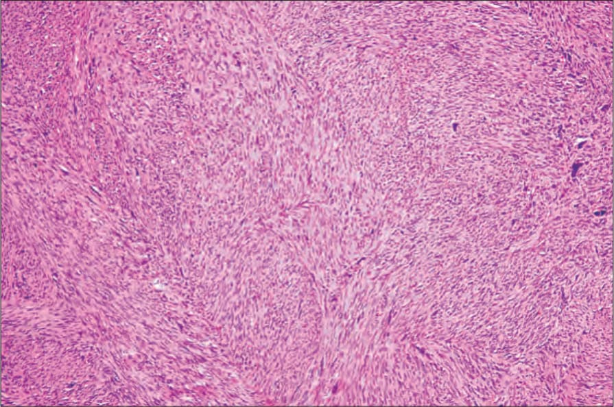
Fig. 12.294 Vulval leiomyosarcoma: this low-power view shows fascicles of tumor cells with eosinophilic cytoplasm. By courtesy of C. Crum, MD, Brigham and Women’s Hospital and Harvard Medical School, Boston, USA.
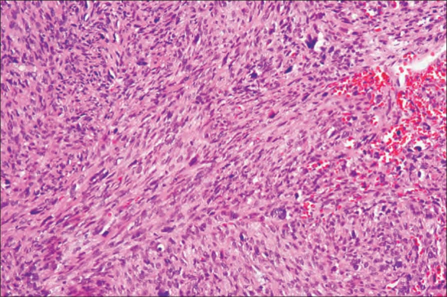
Fig. 12.295 Vulval leiomyosarcoma: note the nuclear pleomorphism. By courtesy of C. Crum, MD, Brigham and Women’s Hospital and Harvard Medical School, Boston, USA.
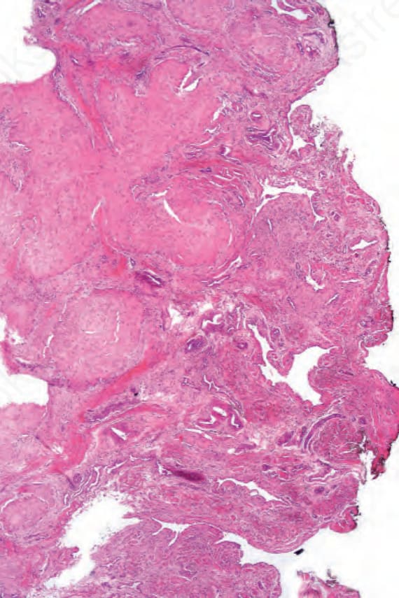
Fig. 12.297 Myointimoma: note the multinodular and plexiform growth pattern.