Angiomyofibroblastoma
Angiomyofibroblastoma
Clinical features This is a distinctive benign soft tissue tumor of the external genitalia and perineum that must be distinguished from aggressive angiomyxoma (see below).1–5 Rare cases have been documented in the vagina, fallopian tube, urethra, and retroperitoneum.6–9 It most commonly affects females of reproductive age but has also been described in the elderly.4 Cases in males are exceptional.10 A tumor sharing many clinical and histologic features with angiomyofibroblastoma has been reported in the male genital tract as angiomyofibroblastoma-like tumor. These are described in the scrotum and groin, and histologically show hybrid features between angiomyofibroblastoma and spindle cell lipoma.11,12
Clinical features Only a handful of cases of this distinctive entity have been reported.1 Patients developed polypoid or cauliflower-like, usually long-standing, lesions on the glans penis or prepuce associated with chronic catheter use and phimosis. All reported cases are in adults with a median age of 40 years. There may be local recurrences.
Histologic features Lesions are polypoid with a hyperplastic epidermis and an edematous stroma with telangiectasia and sometimes focal proliferation of vascular channels. In the background, there are mono- or multinucleated stromal cells and scattered mononuclear inflammatory cells, mainly lymphocytes. The stromal cells are focally positive for actin and desmin.
Angiomyofibroblastoma presents as a slowly growing, small (usually less than 5 cm diameter), asymptomatic subcutaneous mass in the vulva or, less commonly, in the vagina. They are frequently confused with a Bartholin gland cyst. Polypoid morphology is rare.13 but a pedunculated variant is recognized.14 In males, tumors occur on the scrotum and rarely in the perineum, groin, and spermatic cord.15–17 Their behavior is generally benign with little or no tendency for recurrence although a single malignant case has been reported.18,19 This tumor consisted of typical areas of angiomyofibroblastoma with areas of high-grade myxoid sarcoma.
Histologic features Angiomyofibroblastoma is well circumscribed and surrounded by a fibrous pseudocapsule. Scanning magnification reveals a tumor with hypo- and hypercellular areas and a prominent vascular network composed of thin-walled dilated vascular channels (Figs 12.286 and 12.287). The hypocellular areas display prominent myxoid change. Tumor cells are plump, epithelioid, or spindle-shaped with imperceptible to abundant pink cytoplasm, finely dispersed chromatin, and inconspicuous nucleoli. They tend to concentrate around the vascular channels. Multinucleated forms are frequent. Epithelioid cells with hyaline cytoplasm often have a plasmacytoid appearance. Cytological atypia is absent and mitotic figures are usually rare or exceptionally more prominent.20 Scattered lymphocytes and mast cells are often present. Intratumoral mature adipocytes are present in a number of cases and may represent most of the tumor (lipomatous variant of angiomyofibroblastoma).5,21,22 Degenerative nuclear hyperchromatism may sometimes be present.
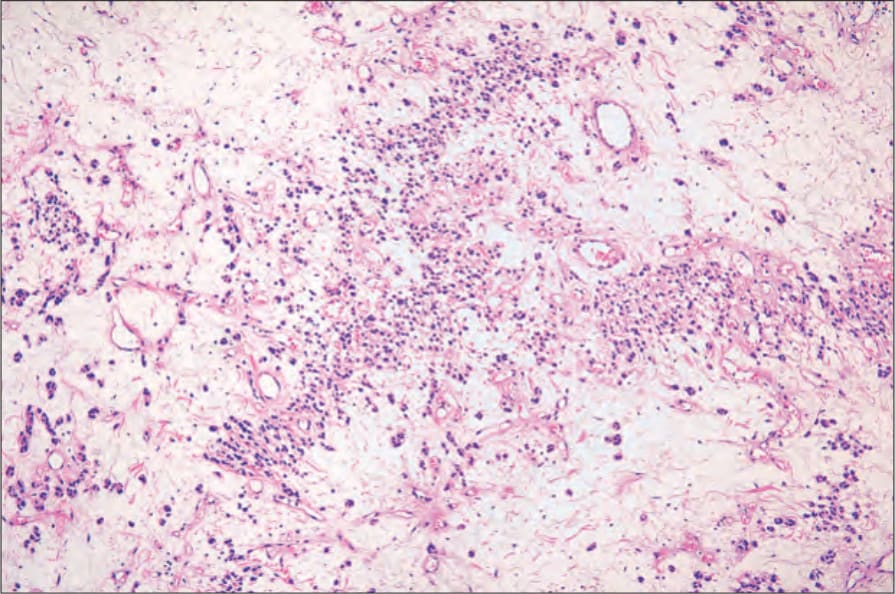
Fig. 12.286 Angiomyofibroblastoma: low-power view showing a richly vascular tumor. In this example, there is a strikingly myxoid stroma. By courtesy of M. Nucci, MD, and C.D.M. Fletcher, MD, Brigham and Women’s Hospital and Harvard Medical School, Boston, USA.