Fibroma
Fibroma
Clinical features Fibroma (fibrovascular polyp, fibroepithelial polyp, irritation/bite fibroma) is the most common tumor-like (but non-neoplastic) condition in the mouth. It is usually located at sites of trauma, namely, the buccal mucosa at or near the linea alba, the lateral borders and tip of the tongue, the lower lip mucosa, and the gingiva.1,2 It presents as a fleshy, pedunculated, or sessile dome-shaped nodule that may be mucosal-colored, ulcerated or keratotic (Figs 11.76 and 11.77).
Pathogenesis and histologic features The fibroma is not a true neoplasm but rather a nodule of scar tissue caused by bite trauma.
The fibroma consists of a mass of fibrocollagenous tissue with variable vascularity and usually mild to insignificant inflammation unless there is overlying ulceration (Figs 11.78 and 11.79). On occasion, the lesion may resemble a hypertrophic scar or keloid. The epithelium may be hyperkeratotic, acanthotic or atrophic. If adipose tissue is present, the term ‘fibrolipoma’ or ‘lipofibroma’ is sometimes applied.
Differential diagnosis Sclerotic fibroma (storiform collagenoma) has been reported in the oral cavity, and the collagen in these lesions has a storiform appearance with clefts between hyalinized collagen and stellate or multinucleated fibroblasts with dendritic processes; multiple such lesions are seen in Cowden syndrome. The cells are sometimes positive for CD34.3
415 Giant cell fibroma
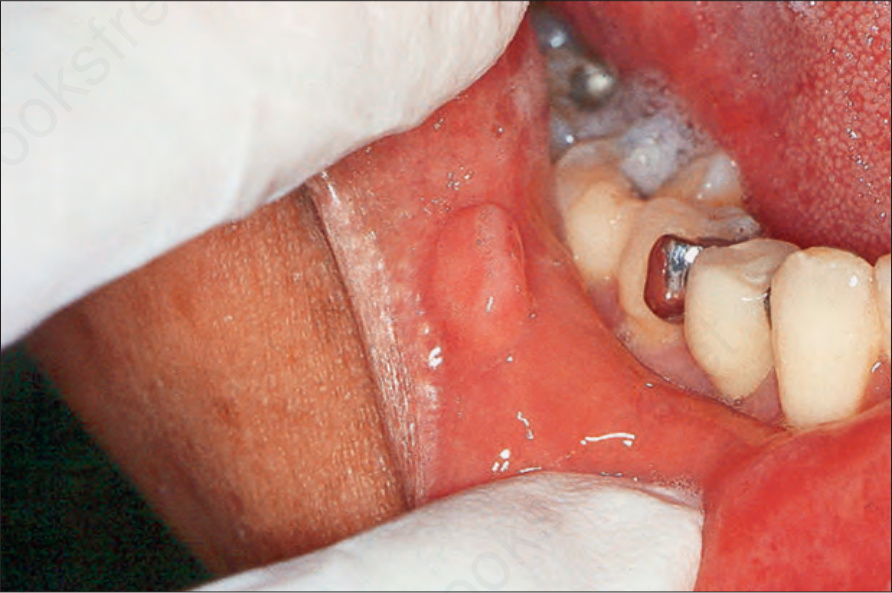
Fig. 11.76 Fibroma: there is a fleshy pink sessile mass on the lower lip mucosa.
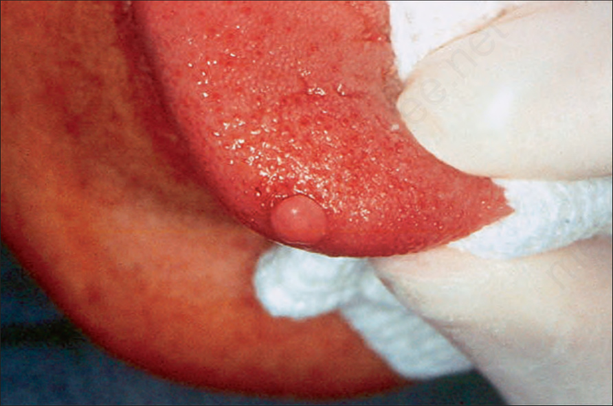
Fig. 11.77 Fibroma: there is a fleshy, polypoid nodule on the lateral border of the tongue.
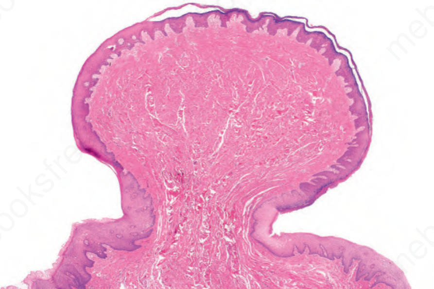
Fig. 11.78 Fibroma: there is a mass of densely collagenous tissue with hyperkeratosis of the epithelium.
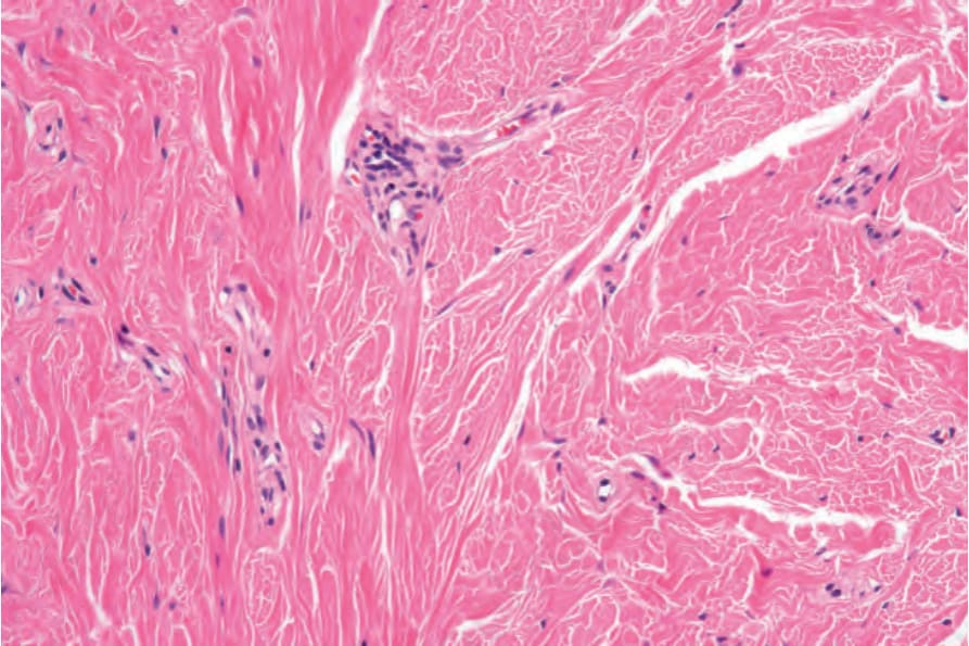
Fig. 11.79 Fibroma: the fibrous tissue is densely collagenous with a few scattered capillaries.
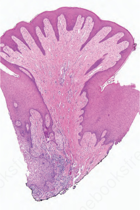
Fig. 11.80 Giant cell fibroma: note the undulating surface and spiky rete ridges.