Leukoedema
Leukoedema
Clinical features
This is a benign, painless condition of adults, usually affecting the buccal mucosa bilaterally, and occasionally the tongue; it is not symptomatic. The mucosa has a diffuse gray-white hue with faint reticulations that disappear on stretching (Fig. 11.29).1,2
leukoedema. The transition zone from normal to degenerate is often abrupt in smokeless tobacco lesion (see below). This should also be considered a severe form of keratinocyte edema caused by contact irritation.
Hairy leukoplakia may exhibit leukoedema and concomitant morsicatio mucosae oris, but will also exhibit viral cytopathic change caused by Epstein-Barr virus.
The prevalence ranges from 20% to 36% among those who do not use tobacco or chew coca leaves, to 51–68% in those who use tobacco, coca, or cannabis.3–8 Its incidence increases with age.9 A prevalence of 51% was reported in African-Americans but without mention of tobacco habits.10 Another study found a prevalence of 93% in a Caucasian population, leading the author to question whether this condition is merely a variation of normal.2
Pathogenesis and histologic features A low-grade topical injury, such as occurs with the use of smoked tobacco or coca leaves, gives rise to this condition. As such, most changes are limited to the superficial layers of keratinocytes.
There is acanthosis with little or no parakeratosis. The characteristic feature is keratinocyte edema where degenerated and swollen cells are present in the superficial and mid-epithelial layers (Fig. 11.30).11 The most superficial cells have abundant pale, watery cytoplasm, sometimes pyknotic nuclei and prominent collapsed cell membranes, forming a jigsaw puzzle pattern; anucleate forms are caused by plane of section of these swollen cells (Fig. 11.31). There is usually no inflammation in the lamina propria.
Ultrastructural studies show abnormal keratohyaline granules and loosely dispersed tonofilaments with fragmented organelles in the superficial degenerated cells. The mid-level swollen cells contain abnormal swollen mitochondria.11,12 These features support the theory of limited cell damage with keratinocyte edema and swelling. Similar features have also been reported in the sucking pads of neonates.13
Differential diagnosis In morsicatio mucosae oris, there is shaggy parakeratosis associated with many bacterial colonies without inflammation; keratinocyte edema is often present beneath areas of such factitial keratosis (see below).
Smokeless tobacco lesion presents with keratin chevrons and shows a band of coagulated and degenerate cells with anucleation similar to
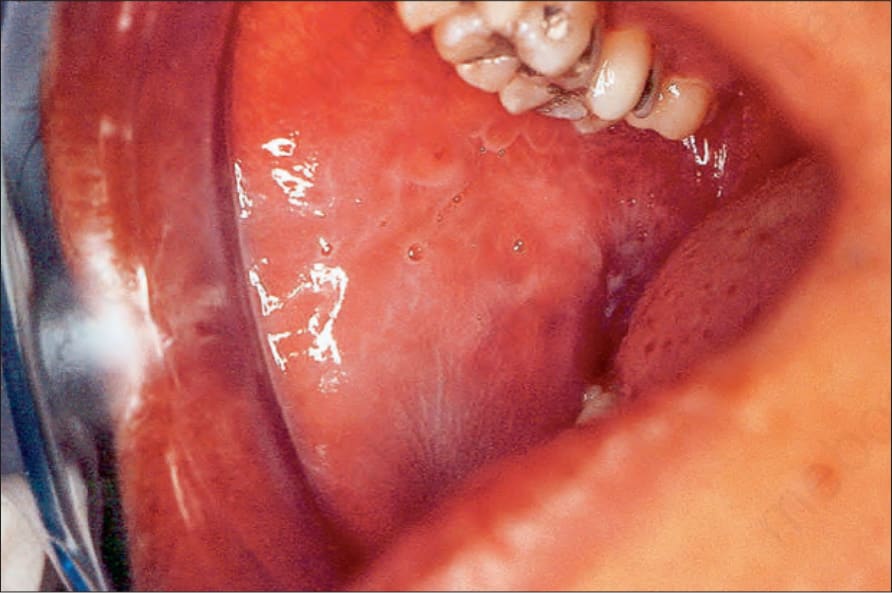
Fig. 11.29 Leukoedema: the buccal mucosa has a pale, milky white appearance.
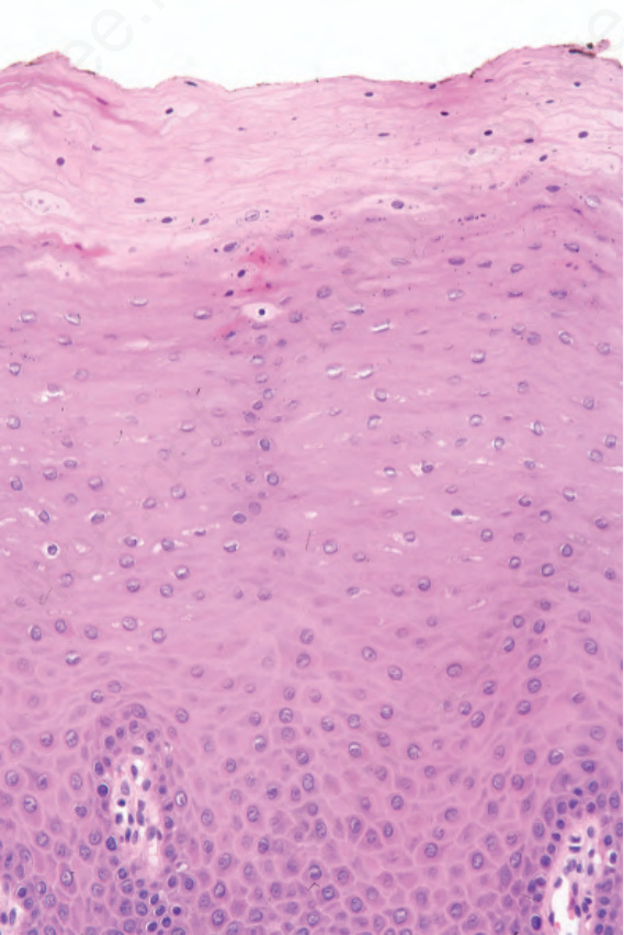
Fig. 11.30 Leukoedema: there is acanthosis and keratinocyte edema of superficial cells.
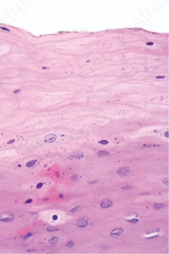
Fig. 11.31 Leukoedema: the superficial cells characteristically have pale cytoplasm and are anucleate with a ‘jigsawlike’ pattern of cell membranes.