Infective granulomata
Infective granulomata
Infections, particularly fungal and mycobacterial, are often associated with granulomatous inflammation. Conversely, the vast majority of routine biopsies showing granulomatous inflammation are not due to an infection. However, when faced with a specimen showing granulomatous inflammation, the pathologist must maintain a low threshold for performing stains for organisms and for issuing recommendations for microbiology culture. If such an approach is not taken, the vast majority of infectious causes of granulomatous inflammation will be misdiagnosed. In addition, although pathologists tend to associate certain patterns of granulomatous inflammation with infection by specific organisms (e.g., caseating granulomatous inflammation and M. tuberculosis), it should be remembered that the same organism can cause several different patterns of granulomatous inflammation (e.g., Mycobacterium leprae). Similarly, mixed suppurative and granulomatous inflammation may result from both atypical mycobacteria and deep fungal infections. A practical corollary to this is that the pathologist should order several special stains to evaluate for a variety of organisms rather than relying on a single stain for the most likely culprit.
338 Granulomatous, necrobiotic and perforating dermatoses
Specific infectious causes of granulomatous inflammation are discussed in detail in Chapter 18.
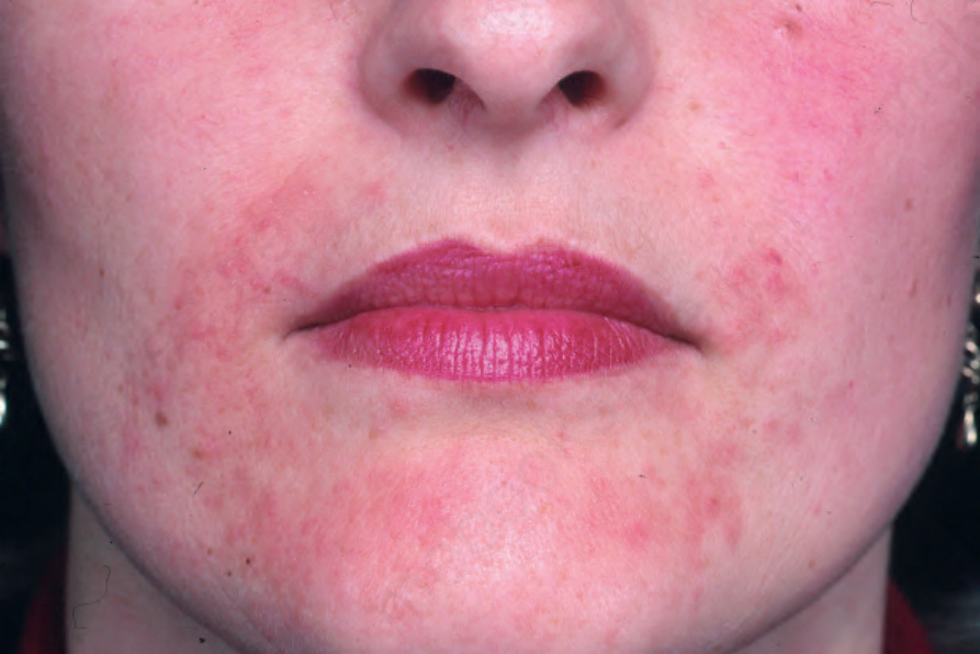
Fig. 9.100 Perioral dermatitis: erythema and papules in a characteristic distribution. From the collection of the late N.P. Smith, MD, the Institute of Dermatology, London, UK.