Elastolytic granulomata
Elastolytic granulomata
This is a controversial group of diseases, the prototype of which is the actinic granuloma. Other entities that very likely belong to this group include atypical facial necrobiosis lipoidica, Miescher facial granuloma, and granuloma multiforme (see below). It has been suggested that all these conditions represent examples of granuloma annulare occurring in different clinical settings.1–4 However, the clinicopathological features are distinctive and the pathological process clearly relates to the primary destruction of elastic fibers by a granulomatous infiltrate. In granuloma annulare, as in diseases such as sarcoidosis, destruction of elastic fibers does not always occur and when it does, it tends to occur focally, developing as a secondary phenomenon.5
328 Granulomatous, necrobiotic and perforating dermatoses
particularly in lesions that do not have an annular configuration. The center of the lesion is completely devoid of elastic fibers, and there is usually no inflammation. The loss of elastic fibers appears to be irreversible.29 In the advancing margin, there is a granulomatous reaction with fragmentation and phagocytosis of elastic fibers. In one case, histology showed features of mid-dermal elastolysis leading the authors to suggest that annular elastolytic giant cell granuloma may possibly represent a prodromal or inflammatory stage of the disease.30 Rarely, sarcoidal granulomas may be a prominent feature.31
Differential diagnosis Many granulomatous reactions, including infections, often display elastophagocytosis. However, in these conditions the change is mild and focal. In granuloma annulare, there is usually very little or no elastophagocytosis, while in annular elastolytic giant cell granuloma there is no necrobiosis, mucin, or palisading granuloma.32 Although a necrobiotic variant of the condition has been described, it is likely that these represent variants of necrobiotic disorders rather than true elastolytic granulomas.9
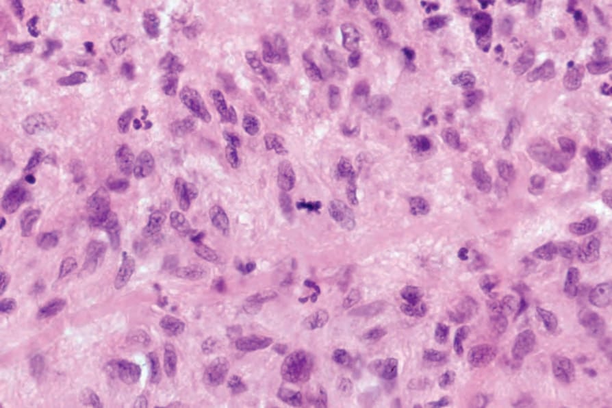
Fig. 9.72 Rheumatoid nodule: the palisading histiocytes may sometimes show mitotic figures which may lead the unwary to consider epithelioid sarcoma.