Granuloma annulare
Granuloma annulare
Clinical features Granuloma annulare is a common, usually asymptomatic, dermatosis of unknown etiology.1,2 It may be divided into six clinical subsets:
• localized,
• generalized,
• perforating,
• subcutaneous,
• papular,
• linear. Unusual clinical variants include pustular follicular lesions and presentation with patches.3,4 A single case presenting as contact dermatitis has been reported.5 Granuloma annulare (often with widespread disseminated lesions) has been described in patients with HIV infection and sometimes may be the presenting sign.6–18 Granuloma annulare, mainly the generalized variant (see below), has also been reported in association with both Hodgkin and non-Hodgkin lymphoma.19–22 Exceptionally, anterior uveitis and concomitant skin lesions have been described.23,24 A case of oral granuloma annulare has been described.25
Other documented associations of granuloma annulare include morphea, chronic hepatitis C infection, autoimmune thyroiditis, secondary hyperparathyroidism, sarcoidosis, Plummer disease, myelodysplastic syndrome, metastatic carcinoma, and a bee sting.26–34 Granuloma annulare has also been described after vaccination for tetanus and diphtheria, hepatitis B, and tuberculosis (BCG), and after mesotherapy.35–39 It may also develop in the scars of herpes zoster.40–43 It is important to highlight that most patients with the condition heal after variable periods of time, and long follow-up has not revealed consistent associations with any systemic diseases.44 A further study has found no consistent relationship between malignant neoplasms and granuloma annulare.45 However, it has been suggested that elderly patients with lesions that do not have typical features of granuloma annulare but display microscopic findings resembling granuloma annulare should be investigated for an underlying malignancy, especially lymphoma.45
the diagnosis of sarcoidosis. In fact, polarizable material has been found in up to 5% of cases.135–137
It has been shown that the gli-1 oncogene is consistently and abnormally expressed in the cells forming the granulomata not only in sarcoidosis but also in granuloma annulare and necrobiosis lipoidica. This observation raises the possibility of trials using inhibitors of gli-1 signaling to treat this group of granulomatous disorders.138
The visceral lesions are characterized by an identical histology of noncaseating granulomata, which may be accompanied by significant scarring, for example, in the lung, where advanced cases are characterized by interstitial fibrosis and sometimes honeycomb lung formation. In the liver, granulomata are most commonly found in the portal tracts or in relation to central veins. Splenic lesions are randomly distributed and are not usually associated with significant fibrosis.
Granuloma annulare has developed during treatment with allopurinol, amlodipine, daclizumab, antitumor necrosis factor agents, thalidomide, vemurafenib, pegylated interferon alpha and topiramate.46–53 Interferon-alpha has been associated with generalized interstitial granuloma annulare.54 It is most likely, however, that granuloma annulare-like eruptions secondary to drug administration often represent interstitial granulomatous drug eruptions.
Differential diagnosis Sarcoidosis must be approached as a diagnosis of exclusion and has to be distinguished from the numerous conditions that may be associated with a noncaseating granulomatous histology, including some forms of tuberculosis, tuberculoid leprosy, berylliosis, fungal infections, Crohn disease, and foreign body granulomatous reactions.139 Therefore, the use of special stains, including the Ziehl-Neelsen preparation for mycobacteria and the periodic acid-Schiff (PAS) and methenamine silver reactions for fungi, is mandatory before diagnosing sarcoidosis. Depending on the clinical context, cultures may also be required to exclude an infective etiology. Tuberculoid leprosy is characterized by nerve involvement, a feature that is usually absent in sarcoidosis.
Localized granuloma annulare The localized variant is the most common type. It usually presents in the first three decades and is associated with a female preponderance (2.25 : 1). Lesions consist of one or several papules, which may be skin-colored, red, or violaceous, and are typically distributed to form an annular or arcuate lesion 1–5 cm in diameter (Figs 9.23–9.27). About 50% of patients have solitary lesions. The acral sites are most commonly affected, in particular, the knuckles and dorsum of the fingers. In a small proportion of patients, lesions are present on both the upper and lower limbs, and occasionally the trunk is affected. Lesions on the palms are exceptional.55 Facial involvement appears to be uncommon.56,57 In a reported case, lesions were restricted to the area involved by a Becker nevus.58 Although lesions may be persistent, approximately 50% of patients can anticipate resolution by about 2 years from onset. However, recurrences are, unfortunately, quite common. Patients in which the disease arises earlier in life appear to have earlier resolution of lesions.59 Interestingly, on occasion lesions regress spontaneously after biopsy.60 Spontaneous resolution may rarely result in mid-dermal elastolysis.61 Rarely, granuloma annulare has been reported in families and in monozygotic twins.62 A case has been documented in which the lesions recurred seasonally with sun-exposed areas.63 There has only been a single case report of cutaneous granuloma annulare with similar lesions in an intra-abdominal location.64 In one case, granuloma annulare was the first sign of adult T-cell leukemia/lymphoma, in another patient it was associated with angioimmunoblastic T-cell lymphoma, and in a further patient it was
Some of the granulomata seen in a variety of primary immunodeficiency syndromes closely mimic those found in sarcoidosis, and histologic distinction may be impossible. A study comparing granulomata in sarcoidosis to those seen in primary immunodefiencies found a much lower rate of CD4+/ CD8+ cells in the former as opposed to the latter.140
Labial and gingival involvement may be histologically mistaken for Crohn disease and granulomatous cheilitis (Miescher). It is worth noting that in rare cases oral involvement in Crohn disease may precede systemic manifestations by several years. Metastatic Crohn disease may be difficult to distinguish from sarcoidosis. The former often shows nonsuppurative granulomata in a diffuse pattern and surrounded by a thin cuff of lymphocytes. Further frequent findings include the presence of numerous eosinophils and ulceration, findings not often seen in sarcoidosis.141
Granulomatous lesions that have been described in exogenous ochronosis appear to be related to sarcoidosis.142 However, similar lesions have also been described as showing changes mimicking actinic granuloma.143
314 Granulomatous, necrobiotic and perforating dermatoses
associated with cutaneous marginal zone lymphoma.65–67 Very rarely, acral, localized granuloma annulare may present as an acute and painful eruption.68 An association with penile lichen sclerosus is exceptional.69
may be asymptomatic or pruritic.2 As with the localized form, the disease is persistent, but some patients experience resolution within 4 years. Anetoderma has been exceptionally reported as a complication of generalized granuloma annulare.75 A remarkable association with giant cell arteritis, gastrointestinal stromal tumor, and other internal malignancies including ovarian and gastric cancer has been reported.76–78 Tuberculous lymphadenits was an association in one case.79 In two instances, the condition was the initial manifestation of chronic myelomonocytic leukemia.80 An association with lymphoma, including Hodgkin disease, has also been described.81 A patient with hepatitis B developed generalized granuloma annulare, and viral DNA was detected in the skin lesions by PCR.82 A further case presented in a photosensitive distribution and healed with scarring and milia formation.83 Other rare associations include hyperlipidemia and scabies.84,85
Generalized granuloma annulare Generalized lesions occur in approximately 15% of patients with granuloma annulare.2,70 As with the localized form, there is an increased incidence in females; however, the median age differs, the majority of cases occurring in patients in the fourth to seventh decades, with the rest appearing during the first decade. Patients with generalized granuloma annulare have an increased incidence of HLA-Bw35.71 Generalized granuloma annulare is defined as lesions occurring on at least the trunk and either upper or lower extremities, or both.2 Most lesions are papules, which may be distributed in an annular pattern, but maculopapules and nodules also occur. They vary in hue from flesh-colored or red, to tan, brown, or yellow. Numbers vary from several dozens to hundreds (Figs 9.28–9.30). A single patient has been documented with generalized disease accompanied by marked swelling of the hands, and another patient developed the disease following erythema multiforme.72,73 A further case developed after varicella zoster infection.74 Lesions
Perforating granuloma annulare Perforating granuloma annulare is distinguished by the presence of transepidermal elimination of necrobiotic collagen.10,18,86–90 Clinically, the lesion presents as a group of papules with an umbilicated crust usually located on the extremities, often the dorsum of the hands (Fig. 9.31). Presentation of
315 Granuloma annulare
lesions on the ears has exceptionally been described, as has a generalized variant.91–93 It may affect both children and adults, and both localized and generalized forms exist. Spontaneous resolution sometimes occurs within months or years of onset. Exceptionally, perforating granuloma annulare develops following tattooing.94
Subcutaneous (deep) granuloma annulare The subcutaneous variant is synonymous with the pseudorheumatoid nodule of childhood and deep granuloma annulare.95–98 Lesions may present de novo or arise in association with typical cutaneous papules. In about a quarter of patients, there is coexistence with dermal granuloma annulare. It occurs in childhood, often affecting the underlying periosteum and involving predominantly the lower legs (particularly the tibia), feet, buttocks, hands, and head.99 Lesions may also present on the penis or eyelid.100,101 An exceptional case of numerous lesions limited to the scalp of a child which regressed spontaneously has been reported.102 A further patient presented with a periorbital subperiostal lesion, and in another patient the lesion was congenital.103,104 In one study of 47 patients, the mean age was 4.3 years.99 In some instances, there is a history of trauma. By definition, such children do not have rheumatoid arthritis or rheumatic fever. The lesion
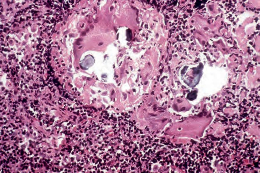
Fig. 9.22 Sarcoidosis: in this lymph node biopsy specimen, fragmented, laminated Schaumann bodies are seen. They are very rarely a feature of cutaneous sarcoidosis.
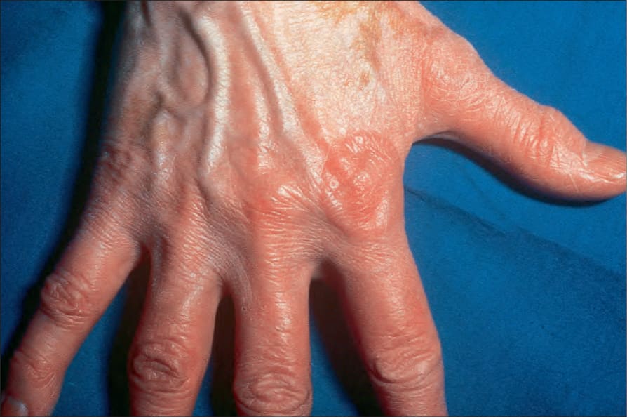
Fig. 9.23 Localized granuloma annulare: a typical annular lesion over the knuckle. Stretching of the skin reveals a translucent beaded margin. By courtesy of R.A. Marsden, MD, St George’s Hospital, London, UK.
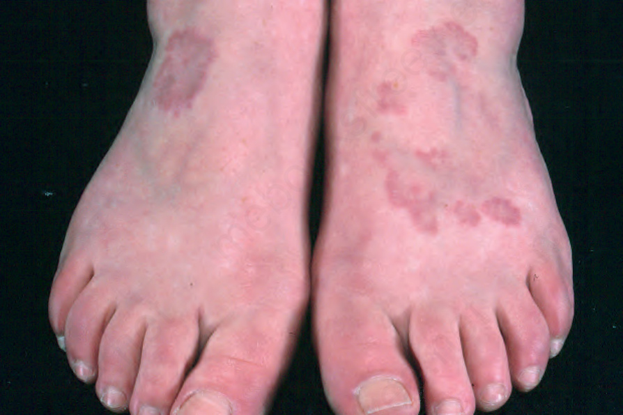
Fig. 9.24 Localized granuloma annulare: in this patient, multiple lesions are present on the feet. From the collection of the late N.P. Smith, MD, the Institute of Dermatology, London, UK.
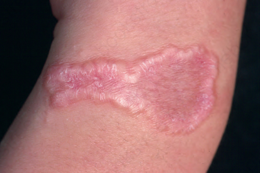
Fig. 9.25 Localized granuloma annulare: this arm lesion shows a characteristic beaded margin. From the collection of the late N.P. Smith, MD, the Institute of Dermatology, London, UK.
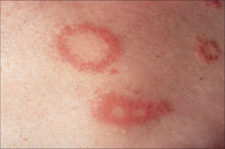
Fig. 9.26 Localized granuloma annulare: close-up view of annular lesions. By courtesy of the Institute of Dermatology, London, UK.
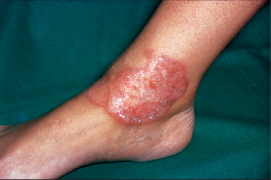
Fig. 9.27 Localized granuloma annulare: in this patient, there is a large plaque on the ankle with a hint of central clearing. By courtesy of the Institute of Dermatology, London, UK.
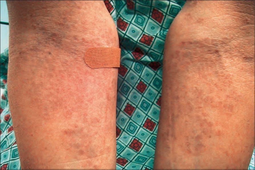
Fig. 9.28 Generalized granuloma annulare: innumerable papules are present on this patient’s arms. By courtesy of J. Williams, MD, Brigham and Women’s Hospital, Boston, USA.
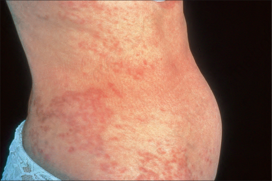
Fig. 9.29 Generalized granuloma annulare: there are widespread papules and plaques. By courtesy of the Institute of Dermatology, London, UK.
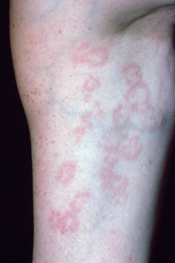
Fig. 9.30 Generalized granuloma annulare: in this patient, numerous annular lesions are present. From the collection of the late N.P. Smith, MD, the Institute of Dermatology, London, UK.
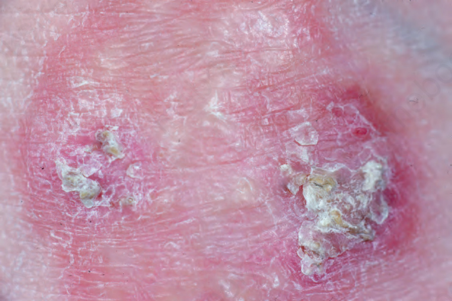
Fig. 9.31 Perforating granuloma annulare: the extremities are most often affected. Necrotic debris and crust can be seen. From the collection of the late N.P. Smith, MD, the Institute of Dermatology, London, UK.
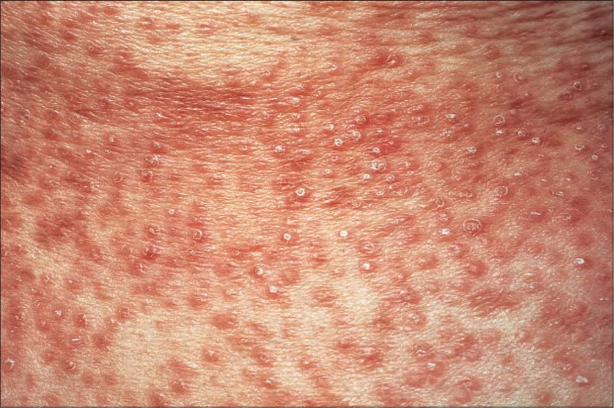
Fig. 9.33 Papular granuloma annulare: numerous small, scaly papules are present. By courtesy of the Institute of Dermatology, London, UK.
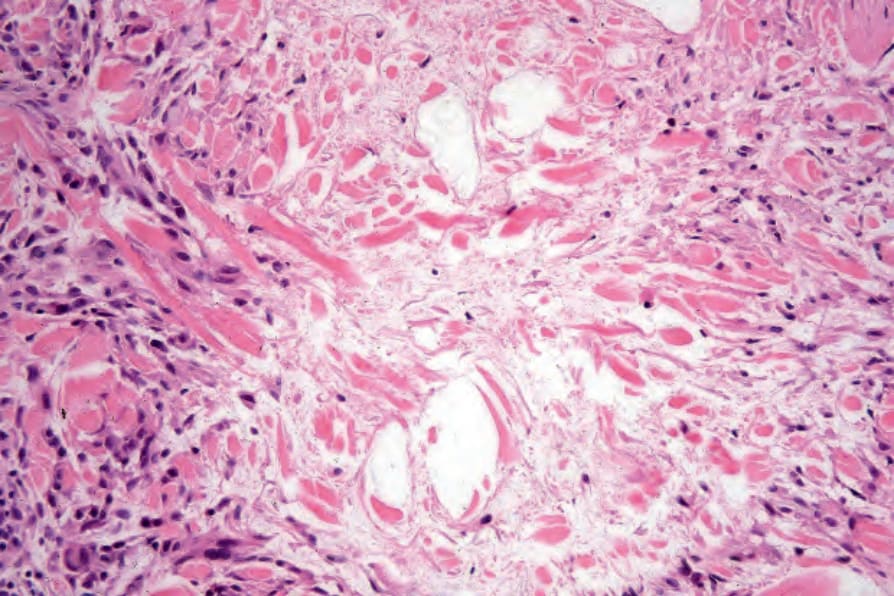
Fig. 9.36 Localized granuloma annulare: the collagen is fragmented and in part granular. Note the peripheral palisade of histiocytes, occasional lymphocytes, and fibroblasts.
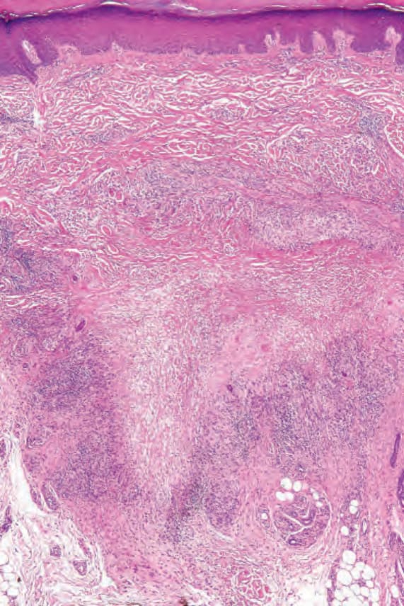
Fig. 9.37 Localized granuloma annulare: this lesion is from the palm of the hand, an uncommonly affected site. There is a sharply delineated focus of necrobiosis in the deep reticular dermis.
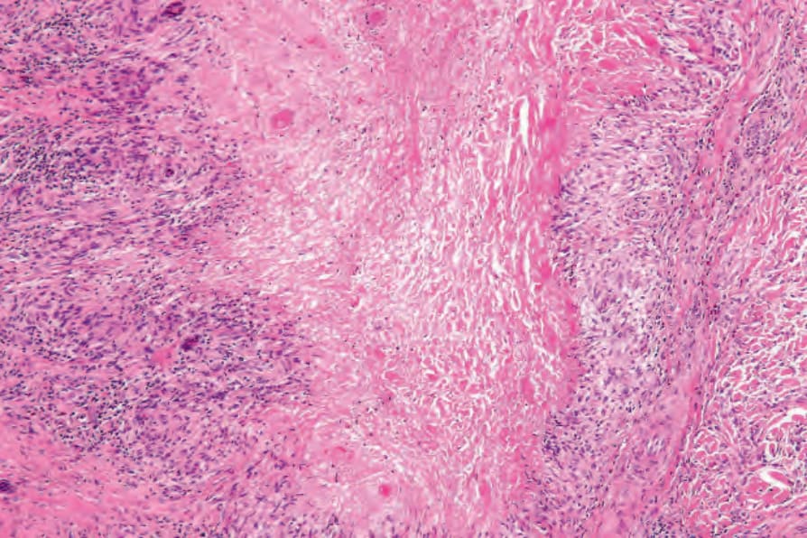
Fig. 9.38 Localized granuloma annulare: the necrobiosis is advanced, presenting as eosinophilic granular debris. The histiocytic palisade is well established.
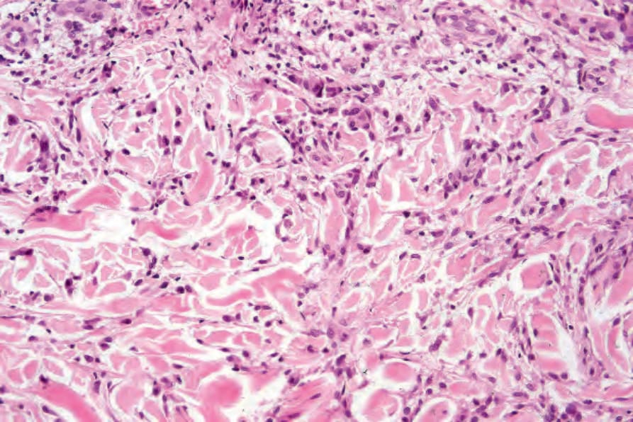
Fig. 9.44 Diffuse granuloma annulare: higher-power view showing the dense interstitial histiocytic infiltrate.
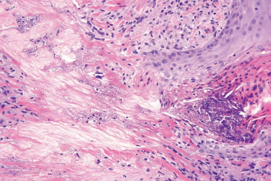
Fig. 9.47 Perforating granuloma annulare: close-up view showing the dermal perforation and transepidermal elimination of collagenous debris. encountered.127 In one study, eosinophils were present in 66% of biopsies, of which 14% showed more than 10 eosinophils per high-power field.126 Plasma cells are rare, and this is useful in the differential diagnosis with necrobiosis lipoidica (see below). Neutrophils are a rare finding and when present, particularly in association with changes of vasculitis, it is likely that there is an association with systemic disease.128 Rarely, an associated prominent lymphocytic infiltrate mimicking a lymphoma may be encountered.129
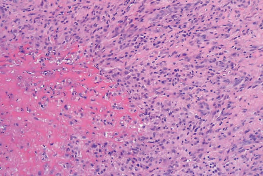
Fig. 9.49 Subcutaneous granuloma annulare: note the intensely eosinophilic necrobiosis and surrounding fibrosis.
usually regresses after several years. However, recurrences appear in 19% of patients.99
Papular granuloma annulare Papular granuloma annulare presents as flesh-colored or hypopigmented, 1–3-mm-diameter papules on the dorsal aspect of the hands, usually in male children. Involvement of the palms and soles may be seen, and rarely lesions are painful.105 Occasional lesions may be umbilicated or generalized (Figs 9.32 and 9.33).106
Linear granuloma annulare The linear variant is very rare and may have a bilateral distribution.107,108 Rarely, it may follow Blaschko lines.109 This variant overlaps and may in
316 Granulomatous, necrobiotic and perforating dermatoses
some cases be the same as the condition described as palisading neutrophilic and granulomatous dermatitid.
Pathogenesis and histologic features The cause of granuloma annulare is unknown. The original concept that it represented a tuberculid has long since been discounted. Although it has been reported at the site of previous herpes zoster infection and verruca vulgaris, it is unlikely that an infectious pathogenesis exists. Borrelia has been demonstrated by focus-floating microscopy in a number of biopsies of patients with granuloma annulare, raising the possibility of a pathogenetic role for the organism in some cases of the disease.110 However, a study based on PCR found no association between granuloma annulare and Borrelia infection.111 There is a wide variety of currently possible pathogenetic mechanisms, most of which have some merit, but none of which satisfactorily clarifies the precise mechanism by which the lesions of granuloma annulare develop.112 Particularly popular are an immune complex vasculitic process and a cell-mediated delayed hypersensitivity reaction. Evidence in favor of the former has been the detection, by direct immunofluorescence, of immunoreactants (IgM and complement) in blood vessel walls in some patients.112 Elevated levels of circulating immune complexes have also been recorded.113 The histology may reveal features suggestive of a vasculitic process, including endothelial swelling, vessel wall thickening (due to the deposition of PAS-positive material), vascular occlusion, and necrosis (Fig. 9.34).114 All of the latter changes may, of course, develop as a consequence of the inflammatory process rather than cause it. A study of serial sections of 38 biopsies in 35 patients found no evidence of a vasculitic process in any of the cases.115
In favor of a cell-mediated delayed hypersensitivity reaction are:
• the finding of activated T lymphocytes in lesions of granuloma annulare on electron microscopic examination,
• the predominance of T-helper inducer cells in the infiltrate,
• the histopathological resemblance of the infiltrate to that of conditions of known delayed hypersensitivity pathogenesis, including sarcoidosis and tuberculosis.116
It has been suggested that Th1 lymphocytes expressing interferon-gamma induce a delayed hypersensitivity reaction leading to macrophages becoming aggressive effector cells that express tumor necrosis factor-alpha and matrix metalloproteinases.117 If tumor necrosis factor alpha plays a role in the induction of the disease, then agents that block this cytokine may be useful in treating the condition. Although some patients respond to these agents, others do not, and the reason for this is not clear. Monozygotic twins with generalized granuloma annulare and the 8.1 ancestral haplotype,
317 Granuloma annulare
a genotype that leads to increased production of tumor necrosis alpha, have responded well to adalimumab.118
Patients with granuloma annulare may have raised serum migration inhibition factor activity.98 Defective neutrophil migration has also been reported.119,120 Other proposed pathogenetic mechanisms include collagen damage by macrophage lysosomal hydrolytic enzymes as the initial event, or a primary disorder of collagen leading to an allergic or nonallergic tissue reaction. The increased incidences of diabetes mellitus and HLA-B8 may also be of pathogenetic significance (compare with necrobiosis lipoidica).121 In a study of a group of pediatric patients with multiple lesions of granuloma annulare, it was found that they had significantly lower serum insulin values than the control group and showed mild impairment of glucose tolerance.122 However, these children often had a family history of diabetes mellitus.
Although there are reports of generalized granuloma being associated with sunlight, this appears to be of doubtful significance.2
It has been shown that the glioma-associated oncogene homologue gli-1, a member of the vertebrate zinc finger transcription factor genes of the gli superfamily, is highly expressed in a number of granulomatous disorders including granuloma annulare.123 The relevance of this in the pathogenesis of granuloma annulare is not clear, but it raises the possibility of using inhibitors of gli-1 signaling in the treatment of granulomatous noninfectious diseases.
The most characteristic histologic lesion seen in granuloma annulare is the palisading granuloma (Figs 9.35–9.38). This consists of a central core of degenerate (necrobiotic) collagen, surrounded by an often radially arranged infiltrate of lymphocytes, histiocytes, and fibroblasts. Elastic tissue may be absent within these foci and there can be phagocytosis of elastic fibers by giant cells at the periphery of the granuloma (Fig. 9.39).124 However, altered elastic fibers are not a constant finding. Solar elastosis is not a feature of granuloma annulare. In some lesions, the altered collagen has a somewhat basophilic appearance due to the presence of acid mucopolysaccharides, but more commonly there is eosinophilia, due in part to fibrin deposition (Fig. 9.40). Heparin sulfate is an important component of the mucin in granuloma annulare but not of other cutaneous diseases associated with mucin deposition (Fig. 9.41).125
Occasionally, sparse karyorrhectic debris is present in the center of the lesion and sometimes the necrobiotic foci contain lipid droplets. More often, however, the collagenous degeneration is not organized into a nodular pattern, but affects isolated fibers in a random pattern, an appearance often best appreciated on low-power examination (Fig. 9.42).126 In this so-called diffuse or interstitial form of granuloma annulare, affected fibers, which are swollen and intensely eosinophilic, alternate with apparently normal fibers
to give a rather disorganized appearance (Figs 9.43 and 9.44). Necrobiosis is minimal or absent. Characteristically, the collagen fibers are separated by mucin, which stains positively with Alcian blue at pH 2.5. Histiocytes are often seen infiltrating around and between affected fibers, and this feature may be a helpful clue to the diagnosis in early cases when the collagen changes are inconspicuous and should, therefore, encourage examination of additional sections to detect more typical features (Fig. 9.45).
An almost inevitable feature of granuloma annulare is the presence of a perivascular chronic inflammatory cell infiltrate, both within the lesion and in the adjacent tissue. Well-formed sarcoidal granulomata with associated giant cells are seen in some cases. Significant numbers of eosinophils may be
318 Granulomatous, necrobiotic and perforating dermatoses
319 Granuloma annulare
are often present. The latter appear to be more common in this variant than in ordinary granuloma annulare. Fibrosis of the surrounding tissue may be marked. In up to 25% of cases of subcutaneous granuloma annulare, changes of classic granuloma annulare may be seen in the dermis.131
Although the relationship with Borrelia infection is debatable, it has been suggested that formation of pseudorosettes in granuloma annulare may suggest infection with the organism.130
In perforating granuloma annulare, the necrobiotic debris is present in close proximity to the epidermis and may be seen to be engulfed by the latter to form a perforating channel by which the necrotic material is extruded to the surface (Figs 9.46 and 9.47). If serial sections are performed, the perforation is often shown to occur through a hair follicle.
The subcutaneous lesions are much larger than the superficial ones (Figs 9.48 and 9.49) and are frequently composed of multiple nodules. There is usually massive necrobiosis and abundant mucin; on occasions, lipid droplets are evident. Mucin, however, may be minimal or not apparent and if fibrin deposition is present, distinction from rheumatoid nodule is impossible. A dense rim of lymphocytes, histiocytes, and fibroblasts surrounds the necrobiotic center. Multinucleate giant cells are common and eosinophils
Papular and linear variants show histologic features similar to those described for typical granuloma annulare.
Differential diagnosis Granuloma annulare must be distinguished from necrobiosis lipoidica, rheumatoid nodule, actinic granuloma, and granuloma multiforme. Points of distinction are summarized in Table 9.1.
The pattern of adipophilin expression has been reported as useful in the histiological distinction between granuloma annulare, necrobiosis lipoidica, and sarcoidosis. In granuloma annulare, adipophilin staining pattern is both intra- and extracellular in relationship with the histiocytes in the infiltrate. In necrobiosis lipoidica the staining pattern tends to be exclusively extracellular in areas of abnormal collagen, and in sarcoidosis the staining pattern is usually exclusively intracellular within histiocytes.132
Occasionally, granuloma annulare may display epithelioid cell granulomas mimicking sarcoidosis. However, the presence of mucin in the
320 Granulomatous, necrobiotic and perforating dermatoses
Granuloma annulare (GA)
Subcutaneous GA Perforating GA
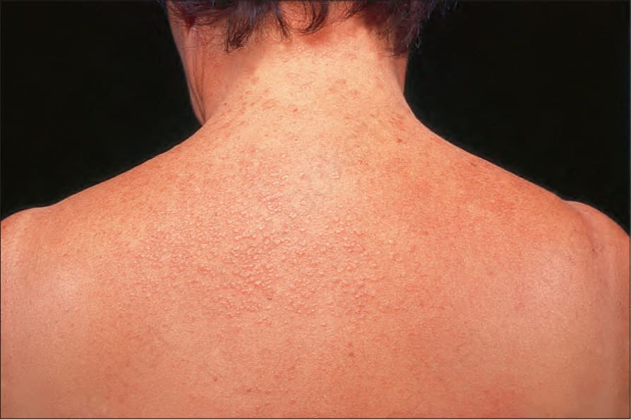
Fig. 9.32 Papular granuloma annulare: widespread papules are present on this patient’s back and shoulders. By courtesy of the Institute of Dermatology, London, UK.
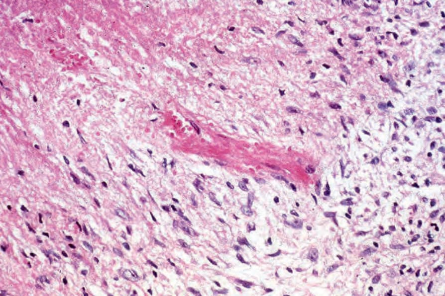
Fig. 9.34 Granuloma annulare: view through the edge of a necrobiotic focus. In the center, a small blood vessel shows fibrinoid necrosis with occlusion. This is an uncommon finding.
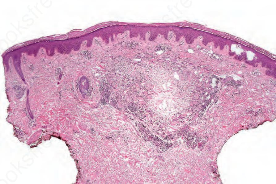
Fig. 9.35 Localized granuloma annulare: the characteristic appearance of a wellcircumscribed palisading granuloma consisting of a necrobiotic center surrounded by a cellular infiltrate.
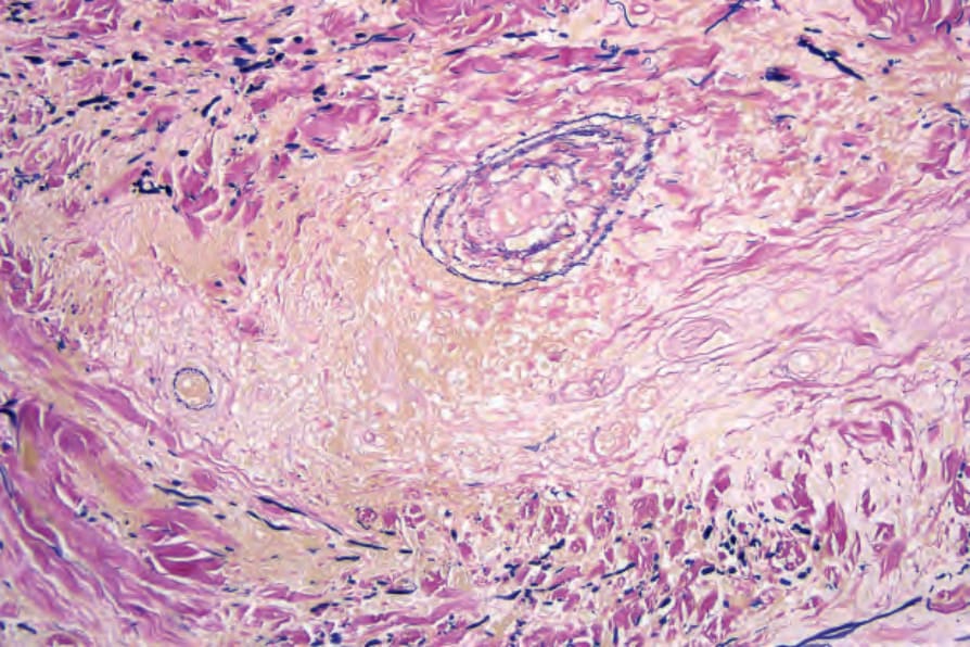
Fig. 9.39 Localized granuloma annulare: there is loss of elastic tissue within the granuloma. Elastic-van Gieson.
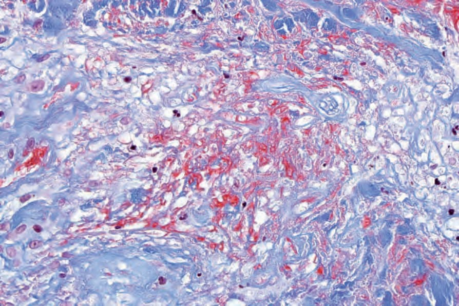
Fig. 9.40 Localized granuloma annulare: the red-staining material in the center of the granuloma is fibrin. Martius scarlet blue.
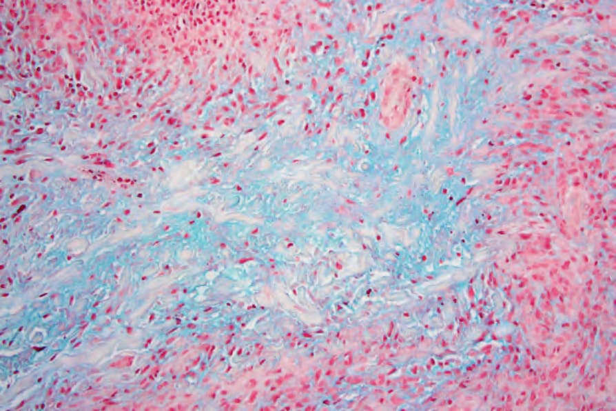
Fig. 9.41 Localized granuloma annulare: in this example, there is abundant mucin in the center of the necrobiotic focus. Alcian blue stain.
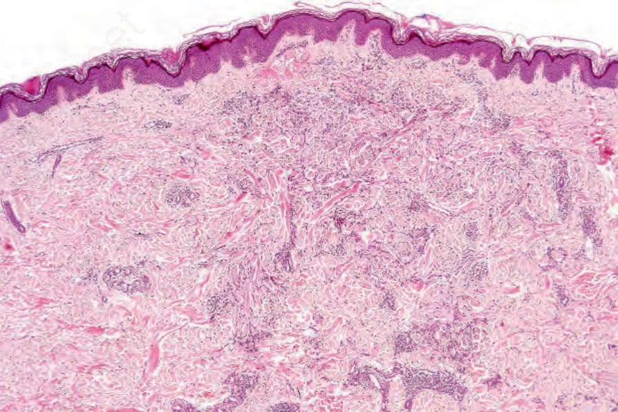
Fig. 9.42 Diffuse granuloma annulare: the collagen bundles are arranged haphazardly. Note the circumferential lymphocytic infiltrate.
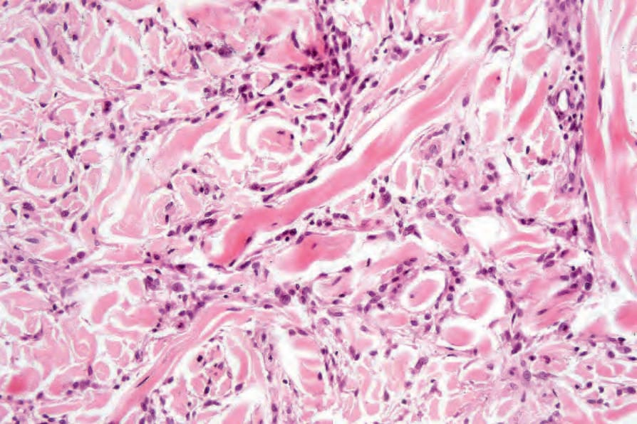
Fig. 9.43 Diffuse granuloma annulare: individual fibers are swollen and intensely eosinophilic. The apparent separation of the fibers is due to increased mucin.
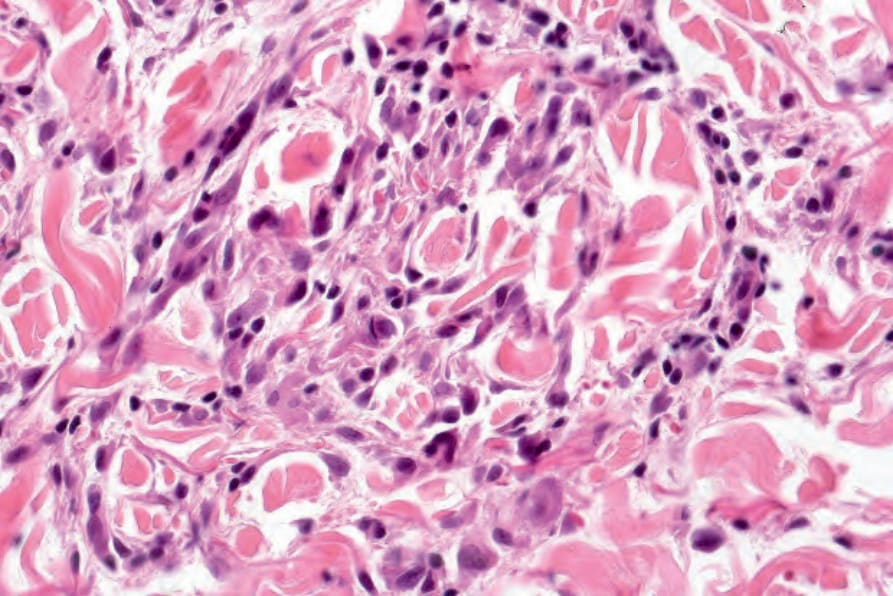
Fig. 9.45 Diffuse granuloma annulare: high-power view.
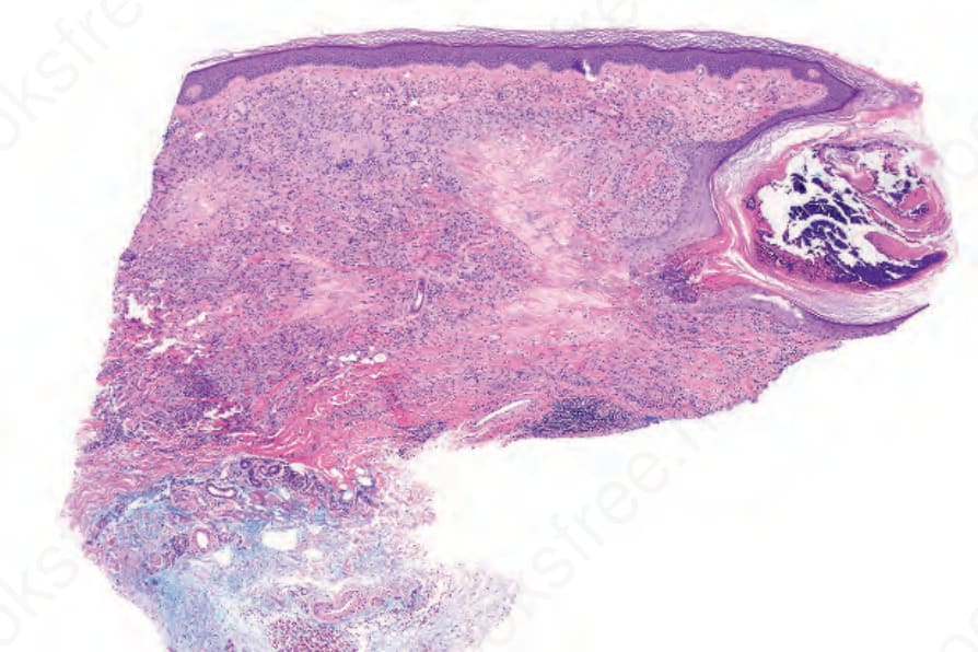
Fig. 9.46 Perforating granuloma annulare: scanning view showing widespread typical granuloma annulare (in the upper-right quadrant, degenerate collagen is undergoing transepidermal elimination).
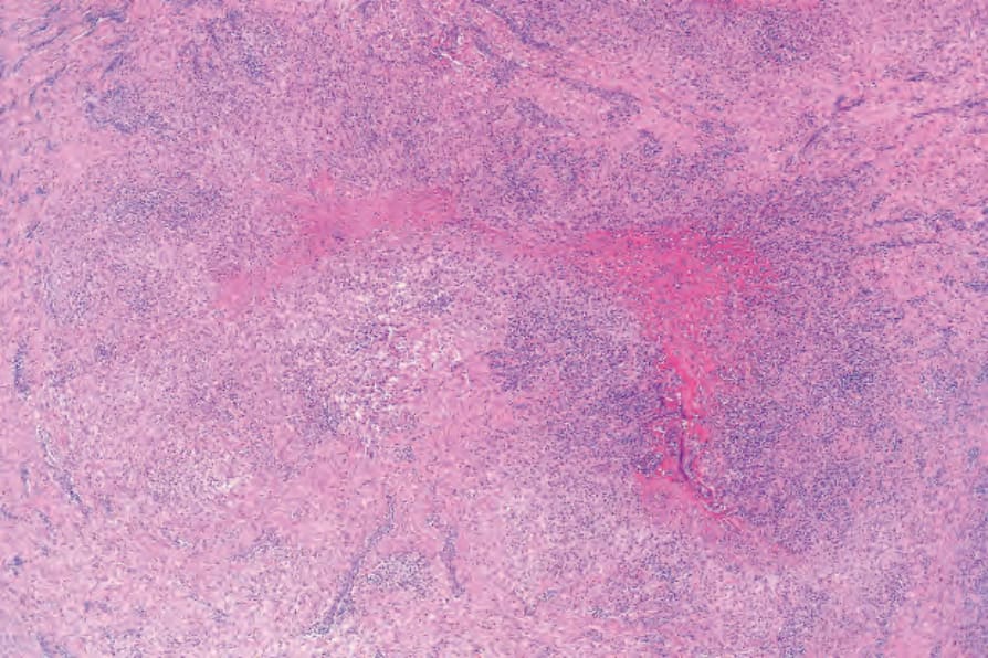
Fig. 9.48 Subcutaneous granuloma annulare: within the subcutaneous fat and involving the fascia is a massive necrobiotic nodule.
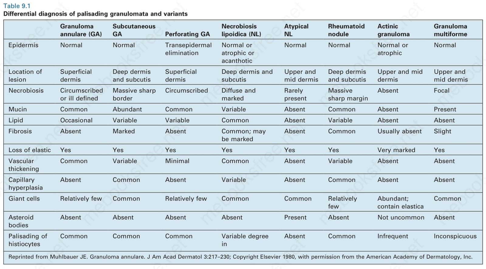
Table 9.1 Differential diagnosis of palisading granulomata and variants
Necrobiosis lipoidica (NL)
Epidermis Normal Normal Transepidermal elimination
Atypical NL
Rheumatoid nodule
Actinic granuloma
Granuloma multiforme
Normal or atrophic or acanthotic
Location of lesion
Superficial dermis
Deep dermis and subcutis
Superficial dermis
Normal Normal Normal or atrophic
Normal
Deep dermis and subcutis
Necrobiosis Circumscribed or ill defined
Massive sharp border
Upper and mid dermis
Deep dermis and subcutis
Upper and mid dermis
Upper and mid dermis
Circumscribed Diffuse and marked
Rarely present
Massive sharp margin
Absent Focal
Mucin Common Abundant Common Variable Absent Common Absent Present
Lipid Occasional Variable Variable Common Absent Variable Absent Absent
Fibrosis Absent Marked Absent Common; may be marked
Absent Common Usually absent Slight
Loss of elastic Yes Yes Yes Yes Yes Yes Very marked Yes
Vascular thickening
Common Variable Minimal Common Absent Variable Absent Absent
Capillary hyperplasia
Absent Common Absent Variable Absent Common Absent Absent
Giant cells Relatively few Common Relatively few Common Common Relatively few
Asteroid bodies
Abundant; contain elastica
Common
Absent Absent Absent Absent Present Absent Not uncommon Absent
Palisading of histiocytes
Common Common Common Variable degree in
Absent Common Infrequent Inconspicuous
Reprinted from Muhlbauer JE. Granuloma annulare. J Am Acad Dermatol 3:217–230; Copyright Elsevier 1980, with permission from the American Academy of Dermatology, Inc.
background along with other more typical changes of granuloma annulare allows distinction in challenging cases.133
Granuloma annulare-like lesions with the added features of vasculitis and a significant component of acute inflammatory cells may be encountered in the setting of systemic disease.129,134 This pattern of disease is discussed in detail in the section on palisaded neutrophilic and granulomatous dermatitis and related disorders.
Granuloma annulare-like drug eruptions have been reported. The presence of associated interface changes favors a drug eruption.10,135
Very rarely, scleromyxedema may focally mimic interstitial granuloma annulare histologically.136 However, the changes simulating granuloma
annulare are focal, and elsewhere in the biopsy there are more typical features of scleromyxedema including fibrosis and increase in fibroblasts.
Occasionally, infection by Mycobacterium marinum may mimic interstitial granuloma annulare. The microscopic features may be so similar that the diagnosis can only be made by special stains and culture.137
Although epithelioid sarcoma, with its associated geographic necrosis, may bear a superficial resemblance at low-power examination to granuloma annulare, the degree of nuclear atypia and pleomorphism in the former condition should afford their distinction in the majority of cases. In addition, epithelioid sarcoma often shows perineural tumor infiltration. It should be noted, however, that mitotic activity may be encountered in granuloma
annulare.138 In difficult cases, keratin, epithelial membrane, in up to 60% of cases, CD34 antigen immunoreactivity and loss of INI1 expression in epithelioid sarcoma should assist in this differential diagnosis.
Rare cases of mycosis fungoides may be associated with a tissue reaction resembling granuloma annulare.139–141 Although interstitial mycosis fungoides may show histiocytes between collagen bundles, the predominance of interstitial lymphocytes with nuclear atypia and epidermotropism, a feature not seen in granuloma annulare, should resolve this differential diagnosis. Most patients with interstitial mycosis fungoides have other classical clinical features of the disease. Of interest, interstitial tumor cells in cutaneous T cell lymphoma often have a cytotoxic phenotype.141
321 Necrobiosis lipoidica
Acrodermatitis chronica atrophicans can rarely display histologic changes resembling granuloma annulare.142 However, clinicopathological correlation and the presence plasma cells in the former usually allows distinction to be made.