Cockayne syndrome
Cockayne syndrome
This is a very rare disorder with an autosomal recessive mode of inheritance and a male predominance (4 : 1), with the majority of cases reported to be of British ancestry. It is a multisystem disease associated with premature aging and particularly affects the skin, teeth, eyes, skeleton, and central nervous system.1
Clinical features Children appear to be normal at birth and have an unremarkable early development. However, usually in the second year of life, they show photosensitivity and acquire a ‘butterfly’ rash (as in lupus erythematosus) on the malar region, which with time is associated with scarring and hyperpigmentary changes. These features, in association with prognathism, sunken eyes, loss of subcutaneous fat, and nasal atrophy (‘beaked’ nose), give the children a characteristic progeria-like or bird-headed appearance (Fig. 7.103).2–4 Fine hair and anhidrosis may also be evident.1
Prenatal diagnosis of Cockayne syndrome is now possible.8
Pathogenesis and histologic features The two genes responsible for Cockayne syndrome (CSA/ERCC8 and CSB/ ERCC6) have been cloned, with most cases due to mutations in CSB.9–12 CSA encodes a WD (Trp-Asp) protein, which interacts with a number of proteins including p44 protein, a subunit of transcription/DNA repair factor IIH (TFIIH).12,13 CSB belongs to the yeast SNF2/SW12 protein family, which is of importance in gene transcriptional activation.12,13 Unlike CSA, CSB is devoid of helicase activity. CSB protein interacts with CSA and excision repair enzyme XPG. It may also have a role in response to hypoxic injury and in chromatin structure.11,12 A mouse model of this syndrome has been developed.14
Ocular lesions include corneal opacity, cataract, retinal degeneration, and optic atrophy with resultant blindness.1 ‘Salt and pepper’ pigmentation of the fundus is characteristic.2
Patients usually suffer from progressive sensorineural deafness.1 The patients are dwarfs and have disproportionately long limbs with enlarged hands and feet.2 Microcephaly is common, and radiological examination reveals thickening of the skull bones. Kyphosis, ankylosis, and flexion
Patients with Cockayne syndrome have an impaired DNA excision/repair mechanism and are hypersensitive to the effects of UV radiation with an inability to promote normal levels of DNA and RNA synthesis following UV irradiation.12,15–18 The specific defect resides within repair of mutations in transcriptionally active genes rather than in excision/repair mechanisms in general.19,20 There are five complementation groups identifiable by cell fusion studies: CSA, CSB, XPB, XPD, and XPG.5,11 XPB, XPD, and XPG differ from groups CSA and CSB by showing an increased incidence of skin cancer.13 Cockayne syndrome may also coexist with trichothiodystrophy.21
Biopsy of the malar rash shows epidermal atrophy associated with basal cell hydropic degeneration. A chronic inflammatory cell infiltrate is present in the superficial dermis.
The cerebral lesions are characterized by loss of white matter, cerebellar cortical atrophy, hydrocephalus, and widespread calcification.5,22 Histologically, there is demyelination and gliosis. Iron-laden neurons, neurofibrillary tangles, and giant, bizarre astrocytes have also been reported.22,23 Severe atherosclerosis resulting in occasional strokes can occur. 22
The kidney shows global sclerosis due to marked basement membrane (type IV) collagen deposition associated with tubular atrophy and interstitial fibrosis.6
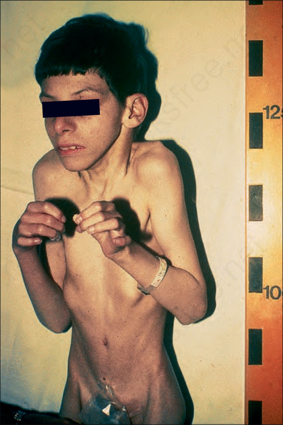
Fig. 7.103 Cockayne syndrome: the features include prominent ears, prognathism, a ‘beaked’ nose, and flexion contractures. By courtesy of D. Atherton, MD, Institute of Dermatology and Children’s Hospital at Great Ormond Street, London, UK.