Definitions
Definitions
There is such considerable variation in the literature as to the exact definitions and interrelationships between erythema multiforme (particularly the ‘major’ variant), Stevens-Johnson syndrome, and toxic epidermal necrolysis that it is often difficult or impossible to be certain to which disease the authors are actually referring!1–5 The consensus paper published in 1993 by Bastuji-Garin is used as a basis for classification because the authorship included most of the major players at that time in this difficult subject.1
reports, the intradermal lymphocytes are also predominantly composed of CD8+ lymphocytes.4,7,12 Immunostains for S100 protein have demonstrated an increased number of Langherans cells within the epidermis.1 Only scattered CD20+ B-cells are present in the dermal infiltrate.
Differential diagnosis The primary differential diagnoses include mycosis fungoides, inflammatory morphea, early lichen sclerosus, and the inflammatory phase of vitiligo. In
Classification of an individual patient depends on the precise morphology and pattern of individual lesions and the extent of skin involvement (detached and detachable epidermis) as a percentage of total body surface area at the worst stage of the illness.
• Target lesions are defined as sharply demarcated and round, less than 3.0 cm in diameter, and comprising three distinct zones, namely, a central erythematous or purpuric disk with or without a blister, surrounded by a raised edematous ring, in turn bordered by an erythematous rim (Fig. 7.68).1 Target lesions are typically distributed in an acral location, are often seen following a herpetic infection, and are characteristic of erythema multiforme. Typical target lesions are not seen in patients with widespread epidermal detachment.
• Raised atypical target lesions are ill-defined, round, palpable lesions with only two zones including a central raised edematous area with an erythematous border.
262 Lichenoid and interface dermatitis
• Flat, atypical target lesions are ill defined, round lesions with only two nonpalpable zones. The center may be blistered (Fig. 7.69).
• Macules with or without blisters are defined as nonpalpable, erythematous, or purpuric macules with irregular shape and size and often confluent. Blisters often occur on all or part of the macule. This lesion is characteristically seen in patients with widespread epidermal detachment who have a history of drug ingestion. Working on this basis, the following definitions have been proposed1:
• Bullous erythema multiforme is characterized by < 10% detachment, typical target lesions, and sometimes raised atypical target lesions.
• Stevens-Johnson syndrome is characterized by > 10% detachment, flat atypical target lesions, and erythematous macules in addition to blisters and erosions affecting one or more mucous membranes.
• Overlap Stevens-Johnson syndrome/toxic epidermal necrolysis is characterized by 10–30% detachment, atypical target lesions, and flat erythematous macules.
• Toxic epidermal necrolysis is characterized by > 30% detachment with flat atypical target lesions and/or erythematous macules. Rarely, toxic epidermal necrolysis may develop as large epidermal sheets in the absence of erythematous macules.
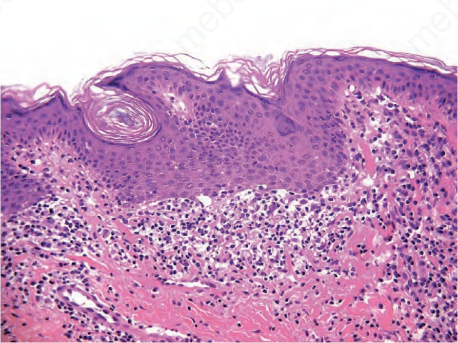
Fig. 7.67 Annular lichenoid dermatitis of youth: the infiltrate causes epidermal necrosis concentrated at the tips of the rete resulting in squared-off, quadrangular rete. By courtesy of Dr. Carlo Tomasini, Torino, Italy.
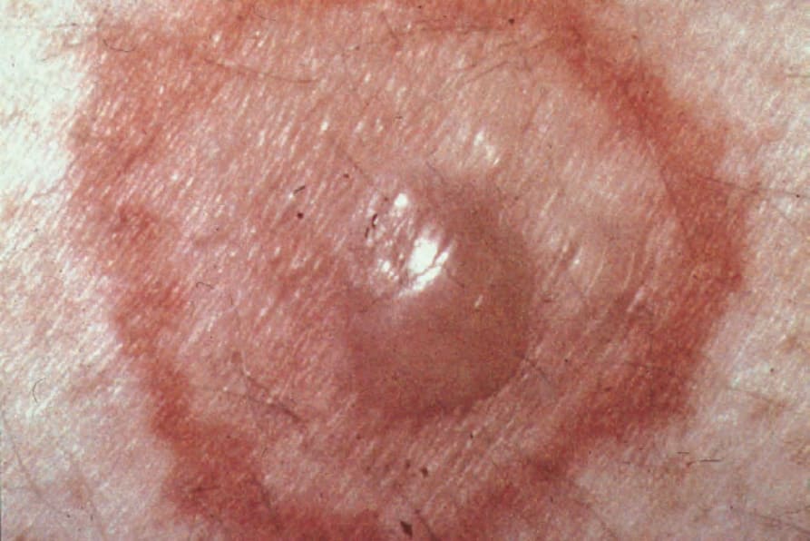
Fig. 7.68 Target lesion: characterized by a central blister surrounded by an edematous ring and an outer erythematous border. By courtesy of R.A. Marsden, MD, St George’s Hospital, London, UK.
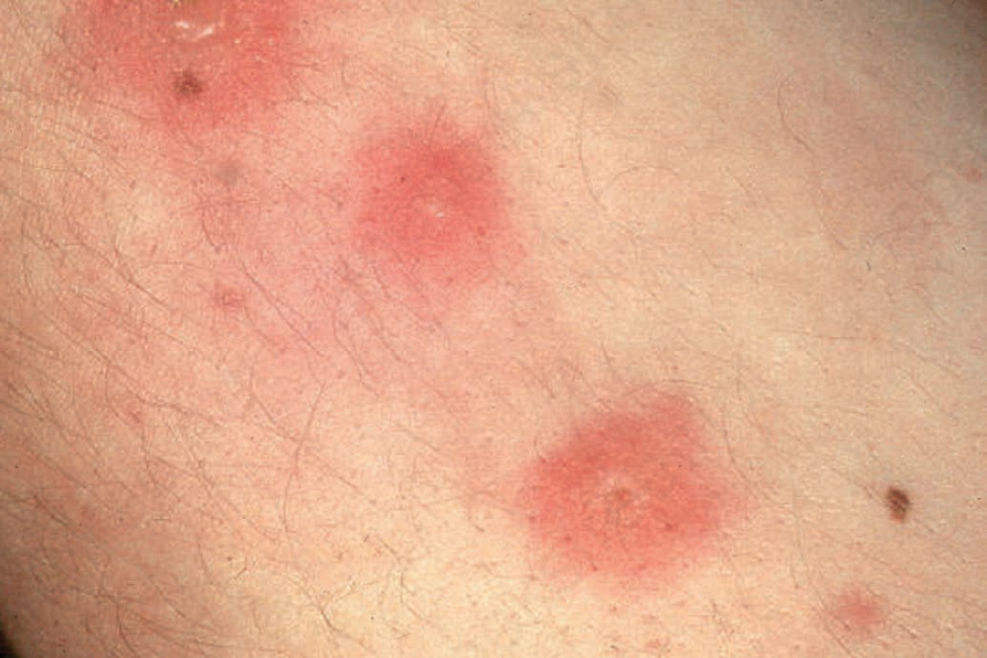
Fig. 7.69 Flat atypical target lesion: characterized by only two components, a central edematous area or blister surrounded by a zone of erythema, these lesions may be seen in erythema multiforme, Stevens-Johnson syndrome, and toxic epidermal necrolysis. By courtesy of the Institute of Dermatology, London, UK.
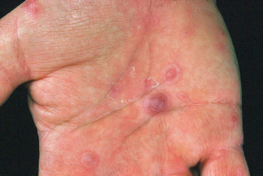
Fig. 7.70 Erythema multiforme: multiple lesions on the hand, a typical site of presentation. From the collection of the late N.P. Smith, MD, the Institute of Dermatology, London, UK.