Acantholytic dyskeratotic acanthoma
Acantholytic dyskeratotic acanthoma
Clinical features Acantholytic dyskeratotic acanthoma is a recently described entity with clinical features similar to acantholytic acanthoma. There is a predilection for the trunk of middle-aged to elderly adults with an equal gender distribution.1 They are solitary lesions characteristically measuring less than 1 cm with a
Pathogenesis and histologic features Focal acantholytic dyskeratosis is a descriptive histopathological term referring to the finding of Darier-like features within the epidermis overlying or adjacent to an otherwise unrelated pathological lesion.1–8 The pathogenesis is not known. The histologic features comprise hyperkeratosis, parakeratosis with suprabasal cleft formation, acantholysis, and dyskeratosis.3 These changes may be seen in the overlying or adjacent epithelium in a variety of lesions, such as basal cell carcinoma, melanocytic nevi, chondrodermatitis nodularis helicis, malignant melanoma, dermatofibroma, condyloma acuminatum, trichofolliculoma, and as part of an epidermal nevus (Fig. 5.82). Focal acantholytic dyskeratosis has been described in a patient with pityriasis rubra pilaris and also in a patient with rosacea.4,8 It has also been described in the oral cavity adjacent to squamous cell carcinoma.9 It is important to recognize this as an incidental finding to avoid misdiagnosis as Darier disease.
Access ExpertConsult.com for the complete list of references
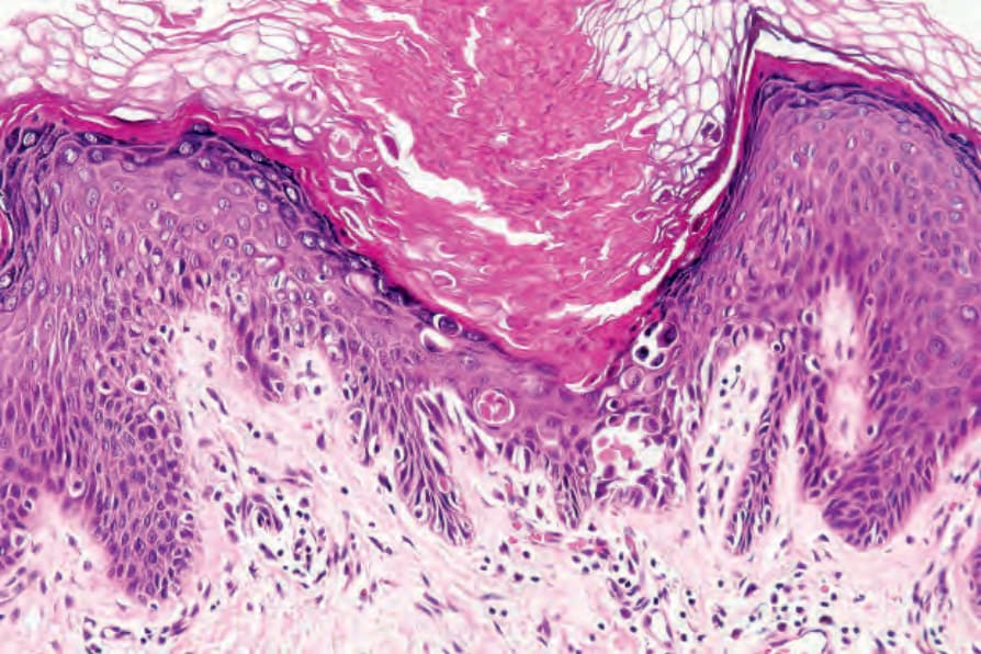
Fig. 5.82 Focal acantholytic dyskeratosis: this example showing the changes of Darier disease was an incidental finding adjacent to a completely unrelated lesion. There was no clinical evidence of Darier disease.