Warty dyskeratoma
Warty dyskeratoma
Clinical features Warty dyskeratoma is a peculiar hyperkeratotic, umbilicated, persistent nodule that usually presents on the sun-exposed skin of the head and neck of middle-aged adults, although lesions on the trunk and extremities, and vulva have been documented (Fig. 5.72).1–5 Most cases are solitary, but occasional patients with multiple tumors have been reported, particularly in Japanese patients.3,6–9 Lesions are commonly asymptomatic but occasionally discharge and bleeding may be encountered.2 There are conflicting data regarding gender distribution in the literature.2,3 Although the cutaneous lesions are believed to be of follicular derivation, histologically similar nodules have been described affecting the oral and vulval mucosa.10–15 The
Histologically, warty dyskeratoma is composed of a widely dilated cup-shaped or cystic lesion containing keratinous debris and often associated with a hair follicle (Fig. 5.73). Superficially, the keratinous debris contains conspicuous corps ronds and grains of Darier. The adjacent and deeper epithelium shows marked acantholysis, and suprabasal villi are a prominent feature (Figs 5.74 and 5.75). The underlying dermis is often infiltrated by lymphocytes and histiocytes, and sometimes plasma cells are evident.
Oral lesions can be morphologically indistinguishable although a number of cases more likely represent focal acantholytic dyskeratosis arising in
198 Acantholytic disorders
a background of a benign trauma-related keratosis. A single case report has documented verruciform xanthoma-like features within a typical oral lesion.13
Differential diagnosis Although there are histologic similarities with familial dyskeratotic comedones, Darier disease, Hailey-Hailey disease, and Grover disease, deeply penetrating crateriform lesions with villus formation are not associated with
these entities. In addition, the clinical findings of a solitary umbilicated nodule should not be confused with any of the above disorders with the possible exception of familial dyskeratotic comedones; however, villi are not conspicuous in the latter. There is also considerable overlap with both focal acantholytic dyskeratosis and acantholytic acanthoma; however, in neither of these conditions is there a deeply penetrating crateriform lesion.
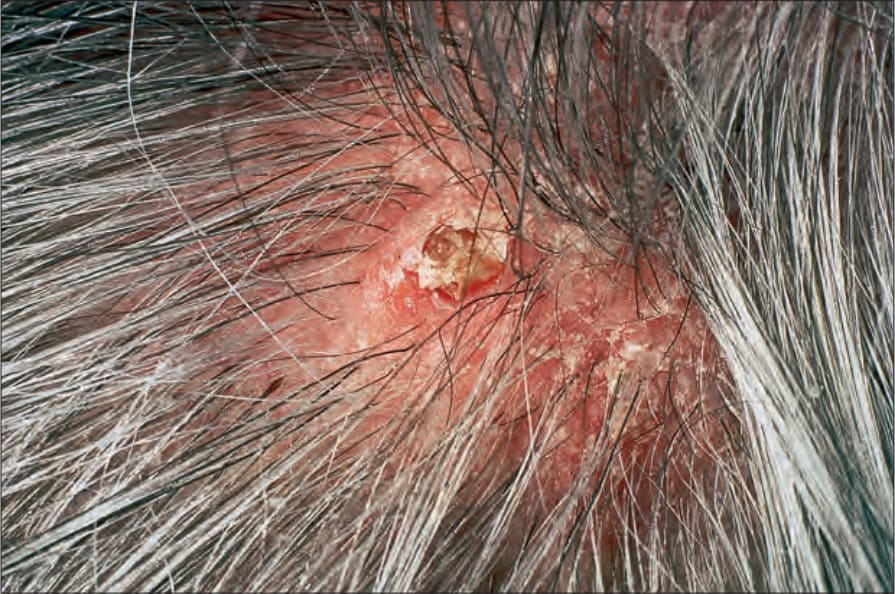
Fig. 5.72 Warty dyskeratoma: scaly nodule on the scalp, a commonly affected site. By courtesy of the Institute of Dermatology, London, UK.
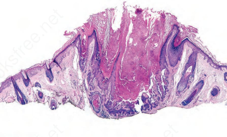
Fig. 5.73 Warty dyskeratoma: typical scanning view of a cystic nodule with acantholysis.
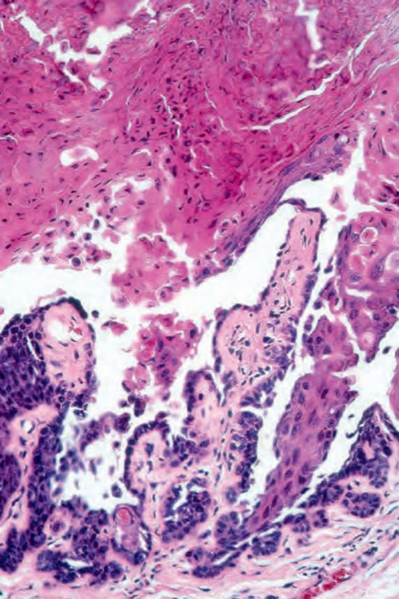
Fig. 5.74 Warty dyskeratoma: note the acantholysis and villi.