Pemphigus herpetiformis
Pemphigus herpetiformis
Clinical features Pemphigus herpetiformis (p. herpetiformis, herpetiform pemphigus, acantholytic dermatitis herpetiformis) is a variant of pemphigus which shows clinical features resembling dermatitis herpetiformis with the histology and immunofluorescent findings of pemphigus.1–7 It is rare, accounting for only up to 7.3% of cases of pemphigus.2,7 The sexes are affected equally, and there is a wide age range varying from newborns to 92 years, although neonatal and pediatric cases are extraordinarily rare.3,7–9
The verrucous plaques and nodules seen occasionally in localized or chronic fogo selvagem show acanthosis, hyperkeratosis, parakeratosis, and papillomatosis.50 Acantholysis is invariably present.
The hyperpigmentation characteristic of remission is a direct result of pigmentary incontinence.
The histologic findings in the endemic form described in the El Bagre area in Colombia are identical to those of fogo selvagem in active disease. In addition, liquefactive degeneration of the epidermal basal cell layer is observed in a quarter of biopsies.28 Patients may also have sclerodermoid changes, psoriasiform features, large subcorneal pustules, and hyperkeratosis of palms.12 By direct immunofluorescence, a positive lupus band test is detected in 40% of patients in addition to IgG deposition on the surface of keratinocytes. Reactive antibodies are of the IgG4 subtype with Dsg1 being the major antigen. Sera from patients also contained additional antibodies against antibasement membrane zone as well as further IgG1 anticell-surface antibodies, which may represent desmoplakin I, envoplakin,
Patients typically present with intensely pruritic, grouped, erythematous papules and plaques, vesicles, and blisters, sometimes associated with mucous membrane involvement.2,7 Urticaria may also be a presenting feature.10 The Nikolksy sign is variably present. Although lesions are often generalized, there is a tendency for the extensor surfaces of the extremities to be particularly involved. Exceptionally, herpetiform pemphigus may be associated with psoriasis, systemic lupus erythematosus, or with an underlying malignancy including lymphoma, lung, prostate, and esophageal cancer, and angiosarcoma (see paraneoplastic pemphigus).7,8,11–16 Although in some patients the clinical manifestations remain herpetiform throughout, in others, the features evolve into more typical p. foliaceus, fogo selvagem, and, less commonly, p. vulgaris.2,4–7 Contrariwise, patients with typical p. foliaceus and p. vulgaris may go on to develop a herpetiform eruption.17
IgA pemphigus may also present with herpetiform lesions.18,19 In general, p. herpetiformis has a benign course, with most patients responding well to sulfones or steroids.2,3,7,20
Pathogenesis and histologic features Immunofluorescence testing shows IgG in an intercellular pattern characteristic of the pemphigus group of disorders on both direct and indirect techniques.1,2,4,7,20 In most patients, Dsg1 (p. foliaceus antigen) is the target autoantigen.4,6,7,21,22 However, in some patients, antibodies against Dsg3 (p. vulgaris antigen) have also been documented.7,22,23 A patient has been reported with both IgG as well as IgA antibodies against Dsg1 in addition to anti-Dsc (desmocollin) 3 IgG.19 Why antibodies to Dsg1 in patients with p. herpetiformis often fail to induce appreciable acantholysis compared with p. foliaceus is uncertain. It is postulated that the p. herpetiformis antibody targets a different epitope although this has yet to be confirmed. Recently, two patients with neutrophil-rich histology were shown to co-localize pemphigus antibody and the neutrophil chemoattractant IL-8. In addition, circulating IgG antibody upregulated cultured keratinocyte IL-8 expression, thereby offering an explanation for the neutrophil recruitment.24,25
183 Pemphigus
The biopsy findings are variable and often non-specific. Although eosinophilic spongiosis is most typical, spongiosis associated with either a mixed eosinophilic and neutrophilic, or a neutrophil-predominant infiltrate may also be encountered.4,26 Intraepidermal vesicles and pustules, also of variable composition, are often present, and dermal papillary neutrophil microabscesses have been described.2,6,20 Acantholytic cells are usually (but not invariably) identified. A requirement for multiple biopsies before a diagnosis can be established is a common theme in the literature.
Differential diagnosis There is both clinical and histologic overlap with IgA pemphigus and dermatitis herpetiformis. Immunofluorescence allows for distinction between these entities. It should also be noted that, exceptionally, dermatitis herpetiformis may histologically show occasional acantholytic cells in the absence of any evidence of pemphigus herpetiformis.
In cases where eosinophilic spongiosis is the predominant histologic feature, the differential diagnosis also includes hypersensitivity reactions and infection (bacterial and fungal). Immunofluorescence studies and special stains for microorganisms will eliminate these possibilities.
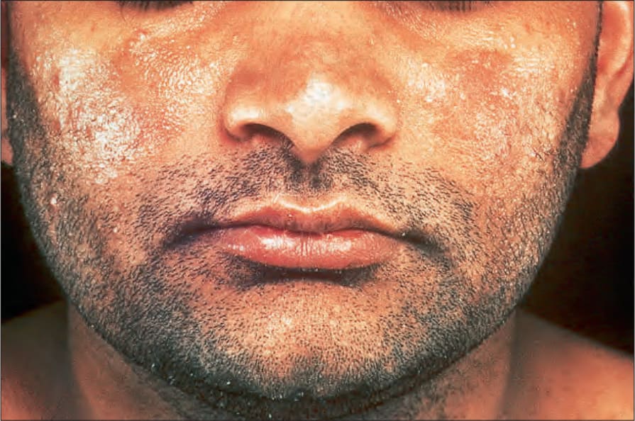
Fig. 5.31 Pemphigus erythematosus: there is scaliness and erythema affecting both cheeks. By courtesy of the Institute of Dermatology, London, UK.