Acantholytic disorders
Acantholytic disorders
for references and additional material
5
Introduction 171 Pemphigus 171 Pemphigus vulgaris 171 Pemphigus vegetans 176 Pemphigus foliaceus 178 Endemic pemphigus foliaceus (fogo
IgA pemphigus 186 Drug-induced pemphigus 187 Contact pemphigus 187 Acantholytic dermatoses with
Transient acantholytic dermatosis (Grover
disease) 195 Acantholytic dermatosis of the genitocrural
area 197 Warty dyskeratoma 197 Familial dyskeratotic comedones 198 Acantholytic acanthoma 199 Acantholytic dyskeratotic acanthoma 200 Focal acantholytic dyskeratosis 200
dyskeratosis 188 Hailey-Hailey disease 188 Relapsing linear acantholytic dermatosis 189 Darier disease 190 Linear Darier disease 194
selvagem) 181 Pemphigus herpetiformis 182 Pemphigus erythematosus 183 Paraneoplastic pemphigus 184
Introduction
The term acantholysis derives from the Greek akantha, a thorn or prickle, and lysis, a loosening. In its simplest definition, the term is used to reflect a primary disorder of the skin (and sometimes the mucous membranes) characterized by separation of the keratinocytes at their desmosomal junctions (Fig. 5.1). A wide range of conditions are characterized by this feature, from inherited disorders such as Darier disease and Hailey-Hailey disease, in which a calcium pump gene mutation results in desmosomal instability, through to the autoimmune pemphigus group of diseases, whereby autoantibodies directly damage desmosomes with resultant keratinocyte separation and blister formation (Table 5.1). Desmosomes may also be damaged by secondary phenomena, for example, following severe edema, either intercellular (spongiosis) or intracellular (e.g., ballooning degeneration as is seen in various viral infections). Such processes, however, are not included in the acantholytic category and are discussed elsewhere. The histologic features of the conditions described in this chapter show considerable overlap. The diagnosis is therefore dependent on adequate clinical information and the results of immunofluorescence investigations.
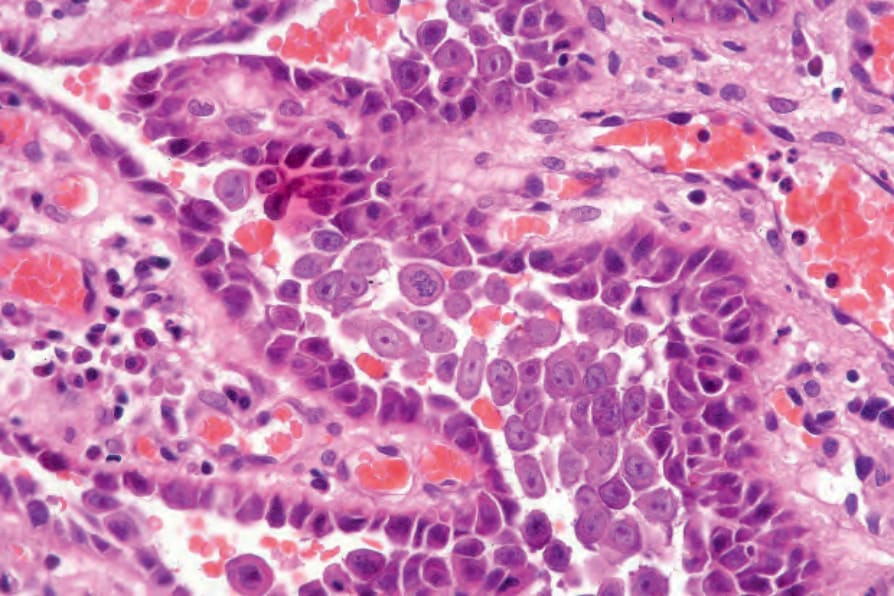
Fig. 5.1 Acantholysis: the keratinocytes are rounded and separated from each other to form an intraepidermal blister. Villi formed from the underlying dermal papillae typically project into suprabasal cavities.
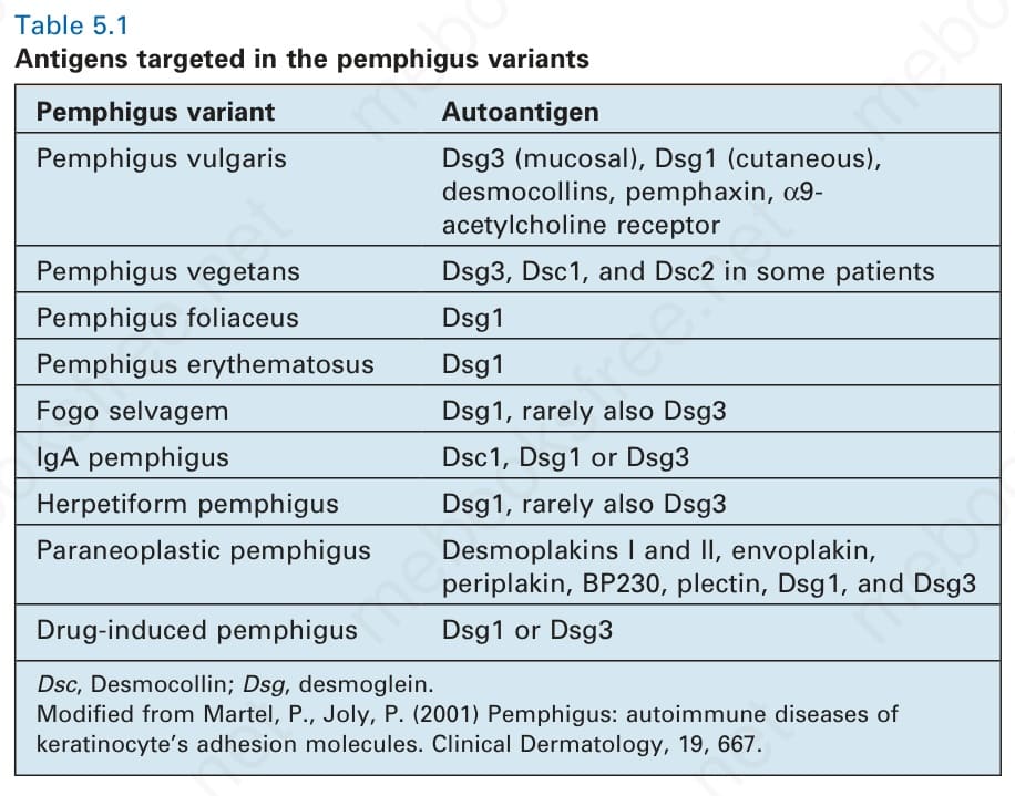
Table 5.1 Antigens targeted in the pemphigus variants