Acquired keratodermas, others
Acquired keratodermas, others
Keratoderma climactericum (Haxthausen disease, climacteric keratoderma) is restricted to menopausal women.1,2 Lesions present on the weight-bearing surfaces of the sole of the foot as erythematous hyperkeratotic and fissured plaques and then spread to involve the rest of the plantar skin (Fig. 3.133). Palmar involvement is sometimes seen with lesions affecting the area between the thenar and hypothenar eminences.2 Similar lesions have been documented in younger women who have undergone bilateral oophorectomy.3 The condition is distinguished from congenital palmoplantar keratoderma by its late onset. Histologically, the plantar skin shows massive hyperkeratosis, hypergranulosis, acanthosis, and spongiosis with lymphocytic exocytosis. A superficial perivascular dermal lymphohistiocytic infiltrate is present and vertically orientated dermal collagen associated with atypical myofibroblasts is often seen.2
Acquired palmoplantar keratoderma can be caused by myxedema associated with hypothyroidism. It improves with treatment.4
Patients with dermatomyositis, in particular dermatomyositis-systemic scleroderma-overlap syndrome may develop circumscribed hyperkeratoses on palms (‘mechanic’s hands’) and soles, sometimes also on elbows and knees (Fig. 3.134). The diagnosis is confirmed by the presence of antibodies against aminoacyl-transfer RNA-synthetase or anti-PM-Scl-antibodies.5 Histologically, subtle signs of interface-dermatitis may be seen.
Chronic lymphedema may also lead to keratoderma. Initially the skin thickens and this is followed by a velvety papillomatous surface, which is ultimately covered by large irregular warty projections.6
Several drugs, such as iodine, lithium, tegafur, and glucan as well as intoxication with arsenic, dioxin, and halogenated weed-killers may also induce keratoderma.7,8 Drugs used for oncologic treatment not only cause palmoplantar erythema, but also keratoderma.9
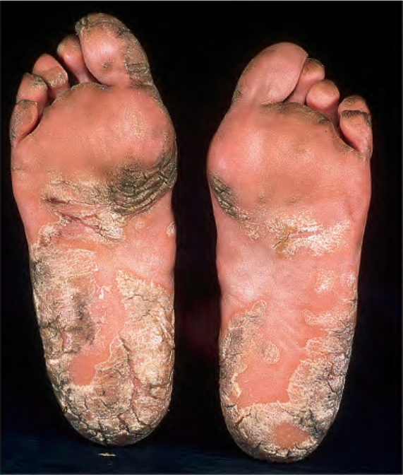
Fig. 3.132 Acquired palmoplantar keratoderma: acquired disease may be a manifestation of underlying malignancy. By courtesy of the Institute of Dermatology, London, UK.
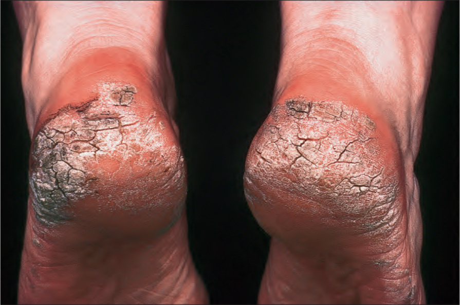
Fig. 3.133 Keratoderma climactericum: there is massive hyperkeratosis with fissuring over the heels. By courtesy of the Institute of Dermatology, London, UK.
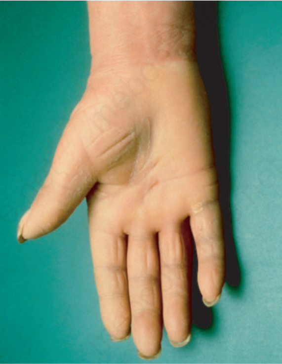
Fig. 3.134 Mechanic hands: patients with dermatomyositis, in particular dermatomyositissystemic scleroderma-overlap syndrome may develop circumscribed hyperkeratoses on palms.