Spiny keratoderma
Spiny keratoderma
Spiny keratoderma is classified as palmoplantar keratoderma punctata type 2 (PPKP2) (syn. music box spine keratoderma, multiple minute palmoplantar digitate hyperkeratosis). In this autosomal dominantly inherited genodermatosis of unknown etiology multiple tiny keratotic spines project from palms and soles including the fingers beginning from the first to third decade of life (Fig. 3.128).1–3 Discomfort is caused by a tendency of the lesions to catch on clothing and other objects.4 Spiny keratoderma has to be differentiated from acquired forms of the disease that evolve after 50 years of age and are associated with neoplastic conditions (visceral carcinoma, melanoma, leukemia, and multiple myeloma) or internal diseases (hyperlipidemia type IV, diabetes, asthma, renal insufficiency, polycystic kidney with liver cysts, tuberculosis, and HIV).2,5–8 Since there is no relation to porokeratosis, old synonyms for spiny keratoderma-like ‘porokeratotic PPK’, or ‘porokeratosis punctata palmaris et plantaris’ should be avoided.
105 Acquired palmoplantar keratoderma and malignancies
Histology demonstrates dense keratotic spikes above a depressed epidermis. Orthokeratosis predominates over focal parakeratotic areas and the underlying stratum granulosum appears attenuated (Fig. 3.129).1,3,4 The absence of dyskeratosis, pleomorphism and vacuolated keratinocytes below the parakeratotic column differentiates spiny keratoderma from porokeratosis and PAON.1 As demonstrated by immunohistochemistry and electron microscopy, keratinization of the keratotic spikes resembles that seen in normal hair cortex suggesting that spiny keratoderma represents an attempt at ectopic hair formation of the palms and soles.9
Degenerative collagenous plaques of the hands affect the sun-damaged skin of the elderly and present as symmetrical yellowish, keratotic or smooth papules and plaques affecting the thumb, first web, and side of the index finger.4,13–18 The ulnar border of the hand and volar aspect of the wrist may also be involved. Keratoelastoidosis marginalis of the hands is a similar condition described in Australians in which keratotic papules develop at sites of trauma along the index finger and thumb.19 The skin is typically grossly sun damaged. Calcified variants of degenerative collagenous plaques are known as digital papular calcific elastosis.20,21
Histologic features Acrokeratoelastoidosis is characterized by massive orthohyperkeratosis overlying a crateriform dell lined by acanthotic epidermis. Hypergranulosis may be present. The dermis shows fragmentation and loss of the elastic tissue (elastorrhexis) (Fig. 3.131). Collagen may be disorganized or appears homogenized and pale staining.2,3
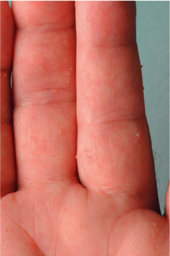
Fig. 3.128 Spiny keratoderma: multiple tiny keratotic spines project from palms, soles, and fingers.
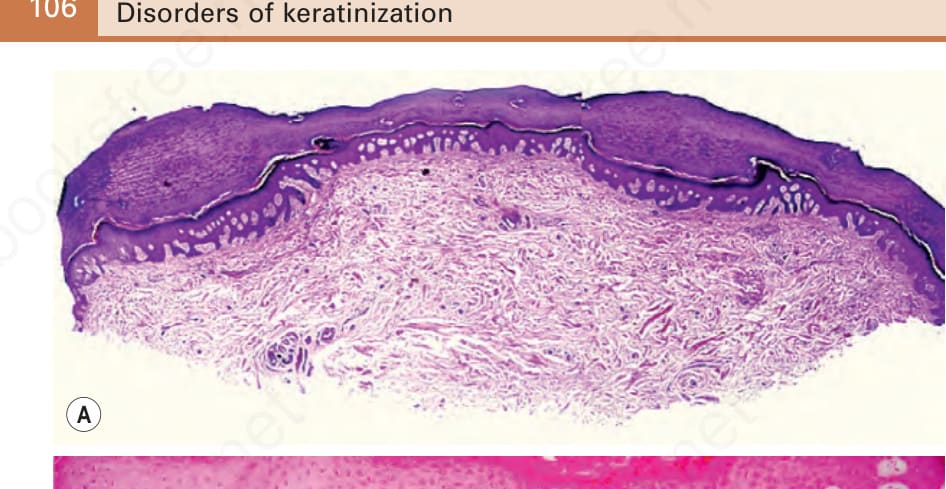
Fig. 3.129 Spiny keratoderma: a keratotic spike develops over a depressed epidermis.
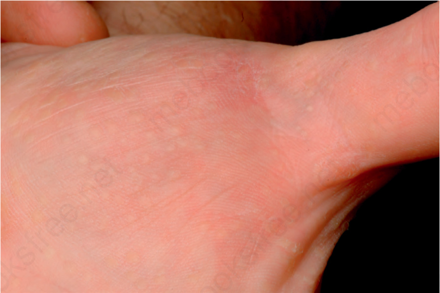
Fig. 3.130 Marginal papular acrokeratoderma: pearly papules predominantly affecting the sides of the hands, wrists, fingers.
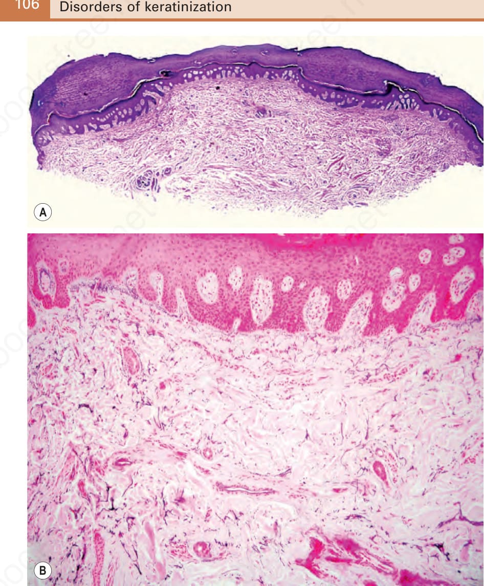
Fig. 3.131 Marginal papular acrokeratoderma, variant acrokeratoelastoidosis: (A) focal areas orthohyperkeratosis overlying crateriform dells lined by acanthotic epidermis; (B) Weigert elastic staining reveals diminution of the dermal elastic tissue.