Striate palmoplantar keratoderma
Striate palmoplantar keratoderma
Clinical features Striate palmoplantar keratoderma (SPPK) is characterized by linear keratosis on palms and island-like areas of hyperkeratosis on soles. Although historically this form of PPK has been regarded as an entity under different designations, such as keratosis palmoplantaris varians Wachters, keratosis palmoplantaris areata et striata, or Brünauer-Fuchs-Siemens syndrome, they are genetically different.1–4 These circumscribed PPKs show an autosomal dominant inheritance and are caused by mutations in at least three different genes coding for desmoglein, desmoplakin and keratin related to keratinocyte adhesion.5
The characteristic clinical features of SPPK are linear keratotic bands on palms and flexor aspects of the fingers and island-like areas of hyperkeratosis
Histologically, striate palmoplantar keratoderma is characterized by massive hyperkeratosis, hypergranulosis, and acanthosis (Fig. 3.114A). Disadhesion of keratinocytes with widening of the intercellular spaces is an important histologic clue in these cell-cell junction diseases (Fig. 3.114B).13 This form of incomplete acantholysis is related to the desmosomal defect and is also seen in Carvajal-Huerta syndrome, McGrath syndrome or diffuse PPK with DSG1 mutation (see Table 3.8).13 At ultrastructural level,
98 Disorders of keratinization
A
the size of the desmosomes appears reduced in DSG1 related SPPK type 1, while perinuclear aggregation of keratin filaments seems more marked in DSP-associated type 2 SPPK.14 Premature expression of involucrin and filaggrin has been described.15
Differential diagnosis The absence of wooly hair and cardiomyopathy clearly rules out Carvajal-Huerta syndrome, another variant of focal or striate PPK (see below). Howel-Evans syndrome must be excluded if focal and nummular keratoderma predominate and histology lacks signs of dehiscence of keratinocytes (see Howel-Evans syndrome).
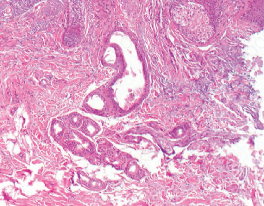
Fig. 3.112 Schöpf-Schulz-Passarge syndrome: the cysts on the eyelids represent apocrine hidrocystomas.
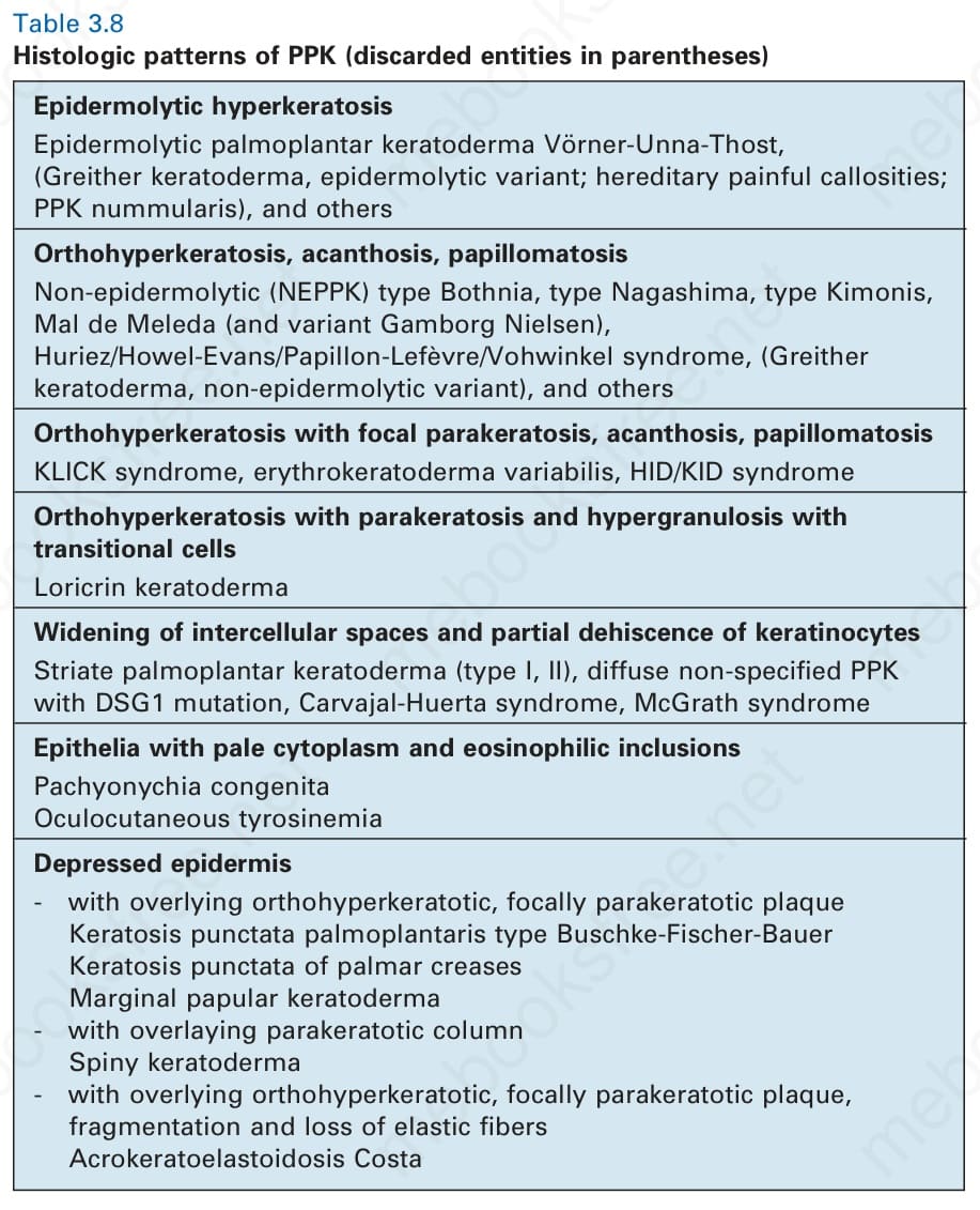
Table 3.8 Histologic patterns of PPK (discarded entities in parentheses)