Erythrokeratoderma variabilis
Erythrokeratoderma variabilis
Clinical features Erythrokeratoderma variabilis (Mendes da Costa syndrome) is a rare ichthyosiform dermatosis with an autosomal dominant mode of inheritance although an autosomal recessive variant has also been described.1–5 Lesions usually present soon after birth or during the first year of life and are of two types, typically occurring simultaneously:
82 Disorders of keratinization
A
B
• Type 1 lesions are symmetrically distributed, discrete figurate, and often bizarre patches of erythema, which vary in size, shape, number, and location over periods of hours and days (Fig. 3.70).3
• Type 2 lesions are well-defined, fixed geographical, reddish-yellowbrown, greasy, hyperkeratotic plaques arising either within the erythematous lesions or, more often, independently (Fig. 3.71). Manifestations of the disease vary within a family and within an individual. The condition affects the face, buttocks, and extensor surfaces of the extremities. Occasionally mild pruritus or a burning sensation are a feature.4 Cold weather in winter, stress, and estrogen-containing oral contraceptives may exacerbate the condition, while the symptoms often improve in the summer months.1,2,4 Hypertrichosis (of vellus hairs) and mild keratoderma of the palms and soles can be present.3,6 The mucous membranes, hair, teeth, and nails are unaffected and there are no associated systemic manifestations.4
a unique clinical feature, namely transient erythematous patches with a peculiar, circinate or gyrate border reminiscent of erythema gyratum repens, i.e., erythrokeratoderma with erythema gyratum repens-like lesions.13–14 Connexin genes code for proteins that form intercellular channels called gap junctions that allow for transport and signaling between neighboring cells in the epidermis. In the skin, Cx31 and Cx30.3 are expressed in the stratum granulosum of the epidermis with a suggested role in late keratinocyte differentiation.12
The histopathological features of erythrokeratoderma variabilis are not specific, consisting of orthohyperkeratosis, variable parakeratosis, irregular acanthosis, and papillomatosis with an undulating skin surface (Fig. 3.72).3,15 Dyskeratotic cells with pyknotic nuclei reminiscent of the grains of Darier have been described (Fig. 3.73).6 The granular cell layer appears normal. A perivascular lymphocytic inflammatory cell infiltrate may be present in the superficial dermis.15,16 Differentiation from psoriasis vulgaris or pityriasis rubra pilaris requires clinicopathological correlation.
Pathogenesis and histologic features In many, but certainly not all families, dominant negative mutations in GJB3 encoding connexin 31 or GJB4 encoding connexin 30.3 have been found.7–10 Autosomal recessive mutations in GJB3 have likewise been reported.11 A subset of patients with connexin 30.3 mutations manifest with
Connexin immunohistochemistry discloses an irregular distribution of the epidermal gap junction proteins in erythrokeratoderma variabilis. Loss of connexin 31 seems to be compensated by connexin 43 overexpression. A cyclic up and down regulation may account for the migratory nature of some lesions. 16
Ultrastructural observations have shown an increased number of gap junctions, some of which display four layers, suggesting a loosened connection of the keratinocyte plasma membrane through the gap junctions.17 Other studies have revealed markedly diminished numbers of Odland bodies in the granular cell layer.6,16 Conspicuous nonmyelinated nerve fibers and Schwann cells have been described in the papillary dermis.6,16 These, however, are not consistent findings.18 Nuclear encirclement by condensed keratin filaments and keratohyalin has also been recorded.18
83 Keratitis-ichthyosis-deafness syndrome, hystrix-like ichthyosis with deafness, porokeratotic adnexal ostial nevus
A
B
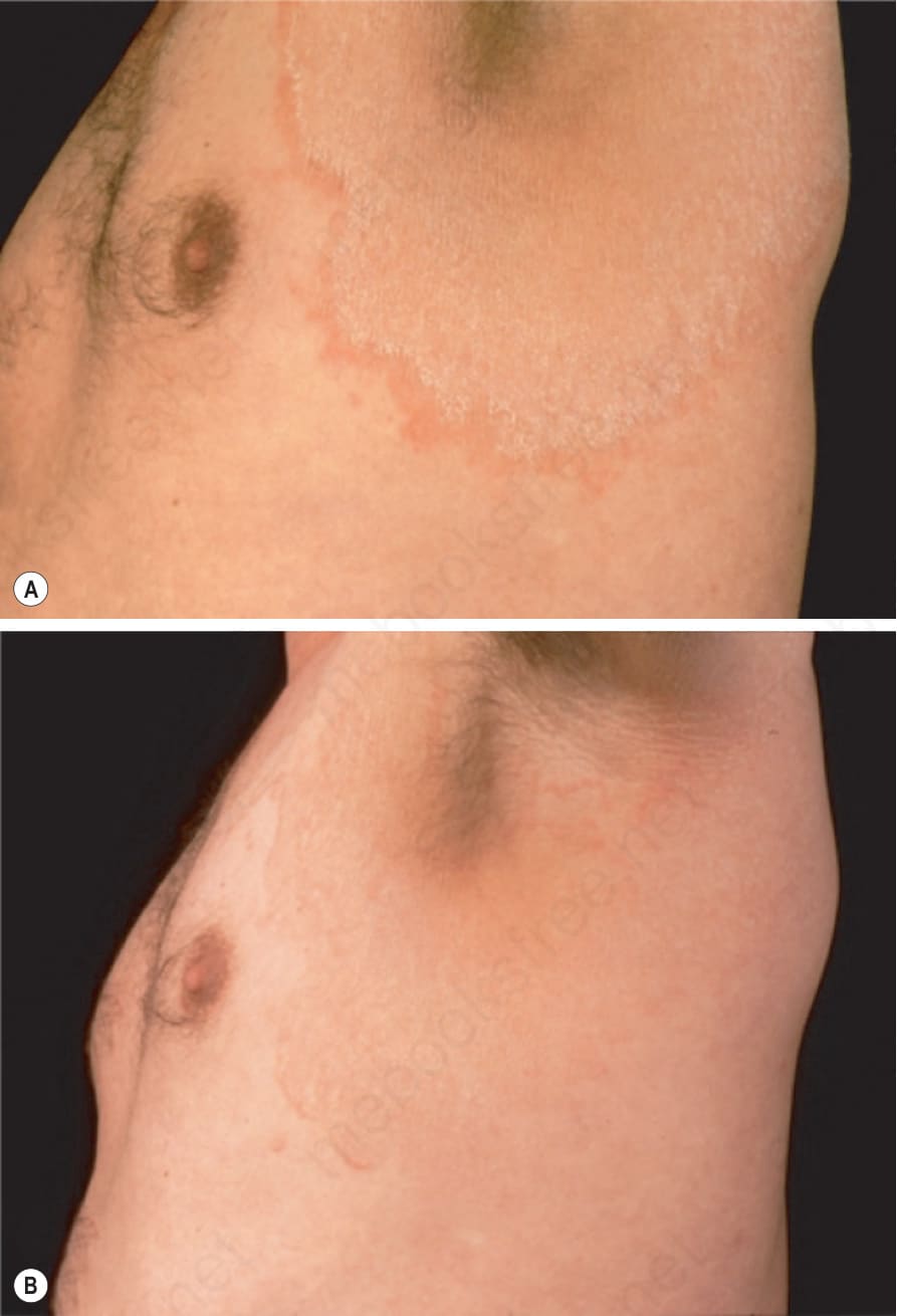
Fig. 3.70 Erythrokeratoderma variabilis: (A) annular erythematous lesions showing scaling; (B) migration within days.
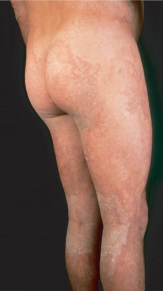
Fig. 3.71 Erythrokeratoderma variabilis: fixed geographical, reddish-yellow-brown greasy, hyperkeratotic plaques.
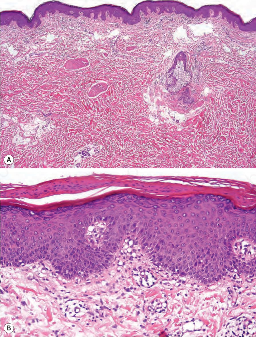
Fig. 3.72 Erythrokeratoderma variabilis: (A) low-power view showing hyperkeratosis, acanthosis with an undulating skin surface and a very mild superficial perivascular chronic inflammatory cell infiltrate; (B) high-power view showing marked parakeratosis overlying a thickened orthokeratotic stratum corneum. Note the presence of a granular cell layer.
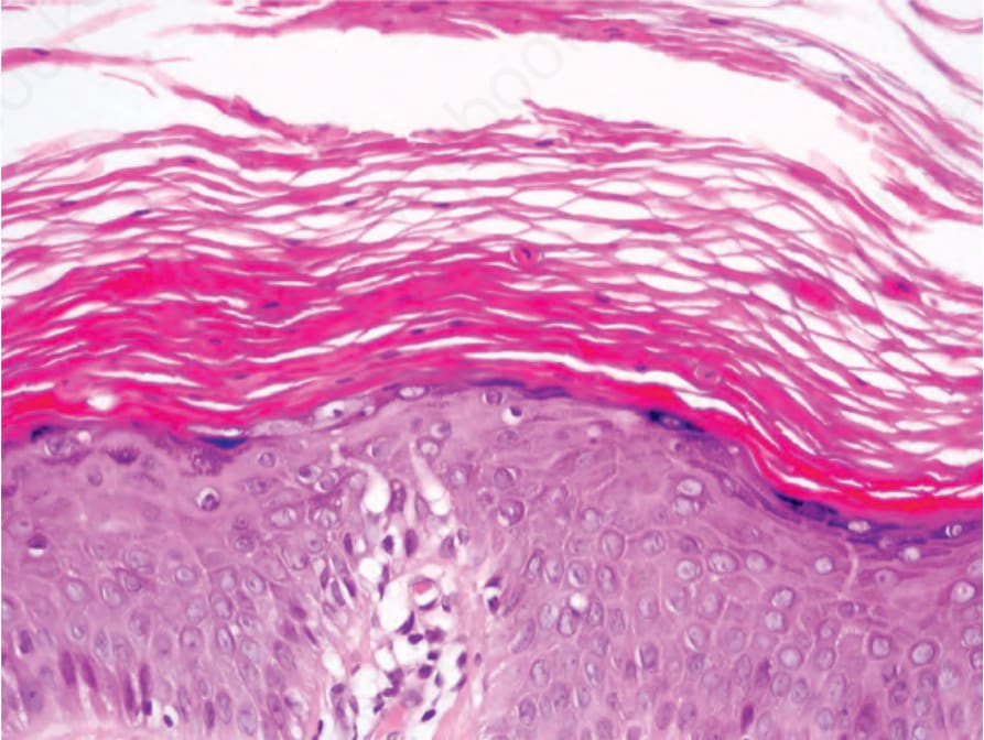
Fig. 3.73 Erythrokeratoderma variabilis: scattered dyskeratotic keratinocytes are sometimes seen.
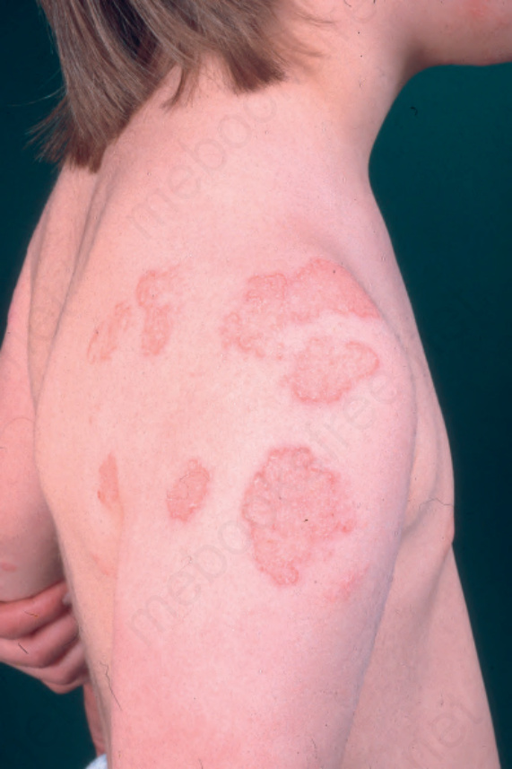
Fig. 3.74 Progressive symmetric erythrokeratodermia: erythematous scaly plaques gradually appear on the extensor surfaces on the extremities and then persist.