Keratosis pilaris atrophicans
Keratosis pilaris atrophicans
Clinical features Keratosis pilaris atrophicans combines the features of follicular hyperkeratosis and atrophic scarring.1 According to different modes of inheritance, clinical presentation, and variable associations three conditions have been described, namely ulerythema ophryogenes, atrophoderma vermiculata, and keratosis follicularis spinulosa decalvans.2
Ulerythema ophryogenes (keratosis pilaris atrophicans faciei, KPAF) presents at birth or in early infancy with follicular papules and surrounding erythema followed by atrophic scarring affecting the lateral aspect of the eyebrows (Fig. 3.67).3–5 The cheeks, forehead, temples, and neck may also be involved (Fig. 3.68). Later on, there may be complete loss of eyebrows. Keratosis pilaris affecting the extensor aspects of the arms and thighs is also
sometimes present.3 The condition is believed to be inherited as an autosomal dominant disorder.
It may be associated with a number of other inherited disorders, including Noonan syndrome, wooly hair, cardiofaciocutaneous syndrome, Cornelia de Lange syndrome, Rubinstein-Taybi syndrome, and partial monosomy 18.3,6–12 The association with Noonan syndrome is of particular importance since such patients suffer from potentially life-threatening congenital pulmonary stenosis. Ulerythema ophryogenes is also associated with atopy.13
Atrophoderma vermiculata (ulerythema acneiforme, atrophoderma vermiculatum, atrophoderma reticulata, acne vermoulante, folliculitis ulerythema reticulata, folliculitis ulerythematosa, honeycomb atrophy) is an exceedingly rare form of atrophic keratosis pilaris with an autosomal dominant inheritance. Patients present with follicular keratoses and pitted depressions separated by normal skin (moth-eaten appearance) affecting the cheeks, ears, and forehead (honeycomb atrophy).2,14–17 The disorder presents
80 Disorders of keratinization
A
B
in patients after 5 years of age.2 Unilateral nevoid variants following Blaschko lines have also been documented.15–17
Keratosis follicularis spinulosa decalvans is characterized by diffuse atrophic keratosis pilaris associated with scarring alopecia affecting the scalp.18–20 Other conditions sometimes present include atopy, palmoplantar hyperkeratosis, photophobia, and punctate keratitis.18 In some patients it is inherited as an X-linked recessive disorder that is caused by mutations in the MBTPS2 gene encoding membrane-bound transcription factor protease, site 2, which has been mapped to Xp21.13-p22.2.21–23 X-linked dominant and autosomal dominant variants have also been described.19 A pustular variant on the scalp that starts at puberty has been described as ‘folliculitis spinulosa decalvans’.24
Pathogenesis and histologic features The pathogenesis of keratosis pilaris atrophicans is unknown. The MBTPS2, as affected in keratosis follicularis spinulosa decalvans and IFAP, may interfere with sterol control and endoplasmatic reticulum stress response.21
All variants of keratosis pilaris atrophicans are characterized by follicular hyperkeratosis with ostial dilatation, atrophy of the sebaceous gland, and a scanty perifollicular and/or perivascular lymphohistiocytic infiltrate. Comedones and milia may be found. There is variable perifollicular fibrosis that extends into the reticular dermis (Fig. 3.69).3,11,12,16
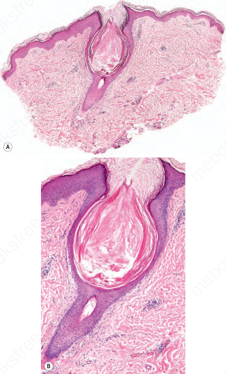
Fig. 3.66 Keratosis pilaris: (A) there is follicular dilatation and plugging; (B) note the atrophy of the infundibular epithelium.
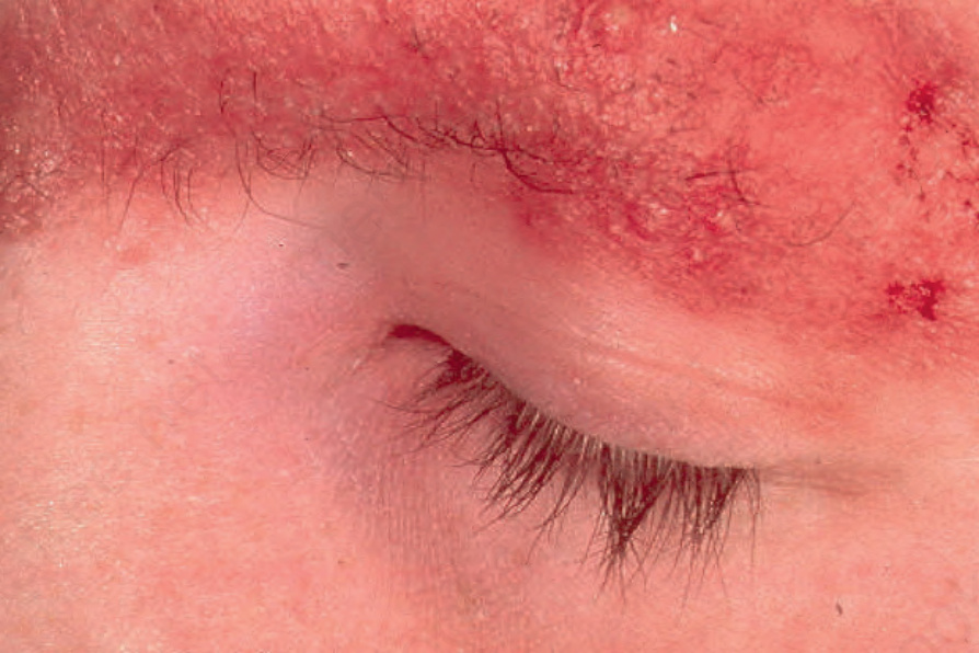
Fig. 3.67 Ulerythema ophryogenes: there is intense erythema with loss of follicles. The eyebrow is a commonly affected site. By courtesy of the Institute of Dermatology, London, UK.
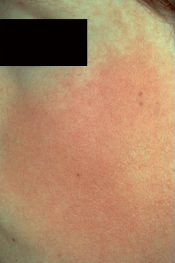
Fig. 3.68 Ulerythema ophryogenes: the cheek is also frequently involved. By courtesy of the Institute of Dermatology, London, UK.
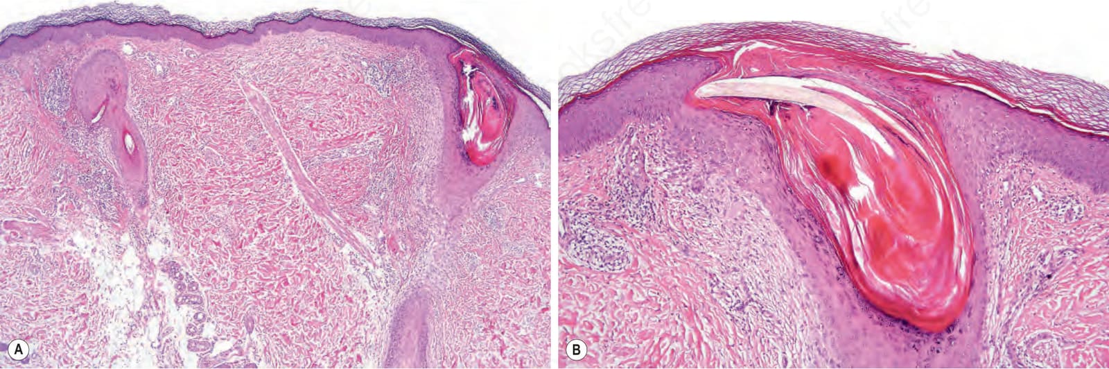
Fig. 3.69 Keratosis pilaris atrophicans: (A) low-power view showing gross follicular hyperkeratosis and dilatation of the ostium; (B) high-power view. Note the perifollicular fibrosis extending into the reticular dermis.