Pityriasis rotunda
Pityriasis rotunda
Clinical features Also known as pityriasis circinata, this acquired disorder of keratinization was originally described in the Japanese.1 It is also not uncommon in South Africans (Bantu) and West Indian blacks,2,3 and has been reported in a subpopulation of Italians in Sardinia.4–7
Patients present with persistent, very sharply defined, circular or oval areas of hyper- or hypopigmentation associated with a fine scale (Fig. 3.64). Lesions, which are usually multiple and frequently numerous, are characteristically noninflammatory and asymptomatic. Often, they are confluent. They measure 0.5–28 cm in diameter and are particularly located on the trunk and limbs. The sex incidence is equal. Lesions are sometimes
78 Disorders of keratinization
A
B
associated with gradual remission during the summer months and relapse in winter.6 The maximum incidence is in the third to fifth decades. There is often a family history of ichthyosis vulgaris.8 Familial cases may occasionally be seen.8,9
Pityriasis rotunda sometimes, but not always, appears to be a cutaneous marker of severe internal disease, including tuberculosis,1 cancer (particularly hepatoma),10,11 myeloma, leukemia,12 cirrhosis,6 ovarian and uterine disease,13 malnutrition, diabetes, and favism.8 Pityriasis rotunda is best be regarded as an acquired circumscribed variant of ichthyosis.12
Histologic features The histologic features are subtle and consist of compact orthohyperkeratosis with a diminished or absent granular cell layer and loss of the epidermal rete ridge pattern. Immunohistochemistry reveals reduced expression of filaggrin and loricrin.14,15 Increased pigmentation of the basal keratinocytes may be evident. A mild perivascular chronic inflammatory cell infiltrate is sometimes present in the superficial dermis. A superficial fungal infection, mainly pityriasis versicolor, should always be excluded.16
79 Acquired ichthyosis-like conditions
A
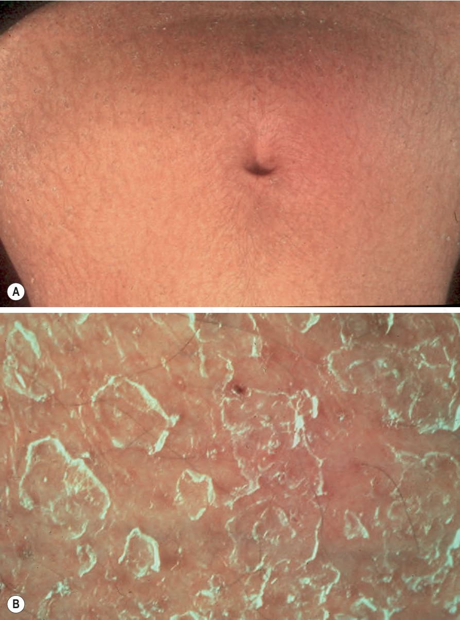
Fig. 3.60 Acquired ichthyosis: (A) cutaneous manifestations most often resemble ichthyosis vulgaris; (B) close-up view of the scale. By courtesy of the Institute of Dermatology, London, UK.
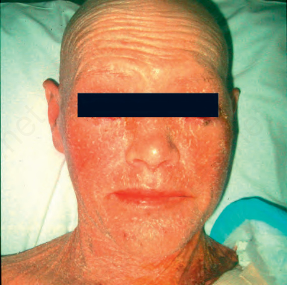
Fig. 3.61 Acquired ichthyosis: there is intense erythema and scaling. This patient also suffered from graft-versus-host disease. By courtesy of B. Solky, MD, Department of Dermatology, Brigham and Women’s Hospital and Harvard Medical School, Boston, USA.
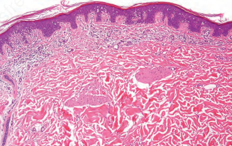
Fig. 3.62 Acquired ichthyosis: this patient developed ichthyosis in a background of mycosis fungoides. Low-power view showing marked focally compact hyperkeratosis and acanthosis.
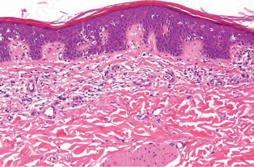
Fig. 3.63 Acquired ichthyosis: high-power view to emphasize the thinned granular layer. Mycosis fungoides as defined by an atypical lymphocyte population and epidermotropism with retraction artifact.
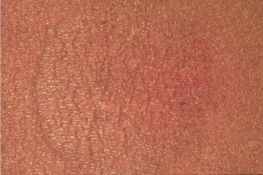
Fig. 3.64 Pityriasis rotunda: characteristic lesion showing circumscription, scaling, and hyperpigmentation. By courtesy of R.A. Marsden, MD, St George’s Hospital, London, UK.