Epidermolytic acanthoma
Epidermolytic acanthoma
Clinical features Epidermolytic acanthoma is an acquired lesion that presents as a verrucous papule or plaque approximately 1.0 cm in diameter and sometimes resembles a viral wart, nevus or seborrheic keratosis.1–3 Caucasians and the Japanese are predominantly affected.3 Lesions may present at any site, but the scrotum, head, neck, and lower limbs are particularly affected.2,3 Although usually solitary, occasional patients may present with multiple localized or disseminated lesions.4–8,11 In Japanese patients multiple depigmented flat keratotic papules following sun exposure on the shoulders and back were described as persistent actinic epidermolytic hyperkeratosis.12 Variants affecting the mucosae of the oral cavity and female genital tract have also been documented.9,10
Pathogenesis and histologic features SEI is associated with a point mutation in the keratin 2 gene (KRT2) located on chromosome 12q11-q13.4–10 Since this keratin is not expressed on volar skin, palmoplantar keratoderma does not develop.
Pathogenesis and histologic features Although it has been assumed that epidermolytic acanthoma develops as a consequence of keratin 1 and 10 gene mutation, two recent genetic studies failed to show any mutations in these keratins.3,13,14 Still, trauma or other external triggers may interfere with the transcription or translation of the suprabasal keratins.13 Human papillomavirus (HPV) infection has been excluded.11
The lesion is well circumscribed and cup shaped and only rarely polypoid. The epidermis shows hyperkeratosis, focal parakeratosis, acanthosis and papillomatosis or a flat base. (Fig. 3.35).1,2 The upper prickle cell
67 Ichthyosis
A
B
and granular cell layers show features of epidermolytic hyperkeratosis, i.e., marked vacuolation of the keratinocytes with eosinophilic keratin inclusions and irregular keratohyaline granules (Fig. 3.36). More variably, eosinophilic perinuclear keratin condensations resembling the Hailey-Hailey pattern of acantholysis are described.11 Necrotic keratinocytes with absent nuclei can be seen in the horny layer. A perivascular lymphocytic infiltrate is often present in the papillary dermis.
Epidermolytic acanthoma displays diminished expression of keratins 1 and 10 and increased expression of the hyperproliferative keratins 6 and 16 while other differentiation markers, such as involucrin and loricrin, appear normal.3,13
are limited to the epidermis overlying just one or two dermal papillae in contrast to the much more extensive involvement in other epidermolytic diseases (Fig. 3.37).
Incidental epidermolytic hyperkeratosis can be associated with epidermal and pilar cysts, scars and fibrous histiocytoma, It may also be seen in normal, particularly sun-damaged skin.1–5 Incidental histopathologic reaction patterns, such as epidermolytic hyperkeratosis, acantholytic dyskeratosis, and Hailey-Hailey-like acantholysis are presumably related to field canceration and can be regarded as a potentially premalignant change surrounding tumors.6,7
Differential diagnosis Identical histologic changes are seen in keratinopathic forms of ichthyosis, linear epidermolytic epidermal nevus, epidermolytic palmoplantar keratoderma, and incidental epidermolytic hyperkeratosis (see Table 3.3). Clinical information is usually necessary to avoid diagnostic confusion.
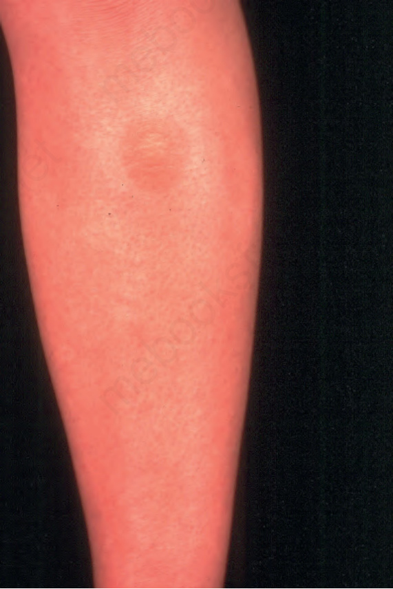
Fig. 3.32 Superficial epidermolytic ichthyosis: flexural hyperkeratosis with early blister formation. By courtesy of W.A.D. Griffiths, MD, Institute of Dermatology, London, UK.
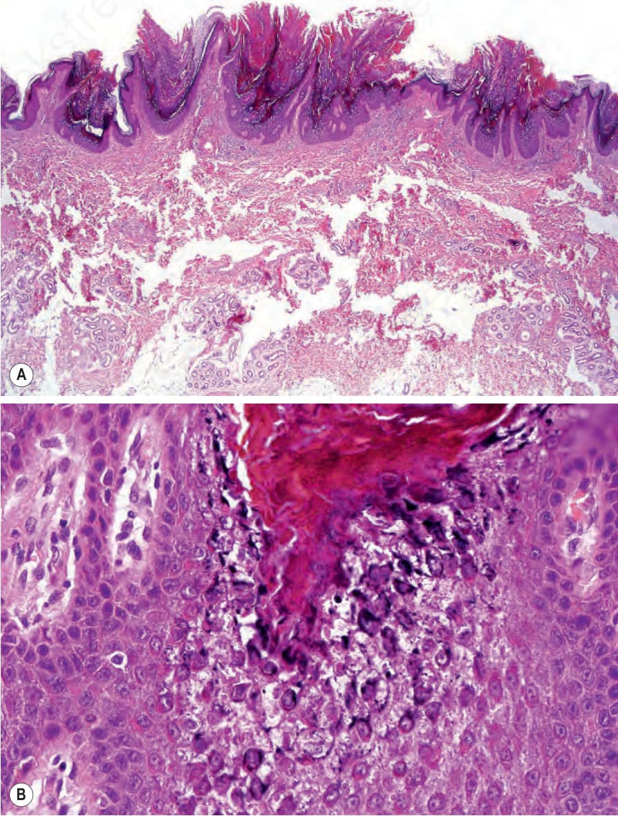
Fig. 3.34 Linear epidermolytic epidermal nevus: (A) low-power view showing massive hyperkeratosis and papillomatosis; (B) high-power view showing epidermolytic hyperkeratosis.
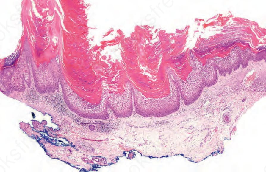
Fig. 3.35 Epidermolytic acanthoma: the lesion is papillomatous with massive hyperkeratosis. There is a superficial perivascular chronic inflammatory cell infiltrate.
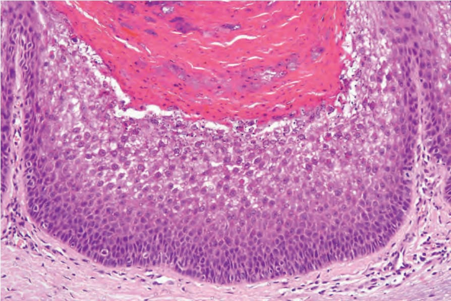
Fig. 3.36 Epidermolytic acanthoma: there is superficial cytoplasmic vacuolation and eosinophilic inclusions are conspicuous.
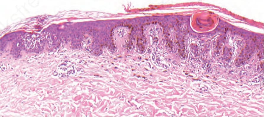
Fig. 3.37 Incidental epidermolytic hyperkeratosis: focal expression of epidermolytic hyperkeratosis in the periphery of a melanocytic nevus.
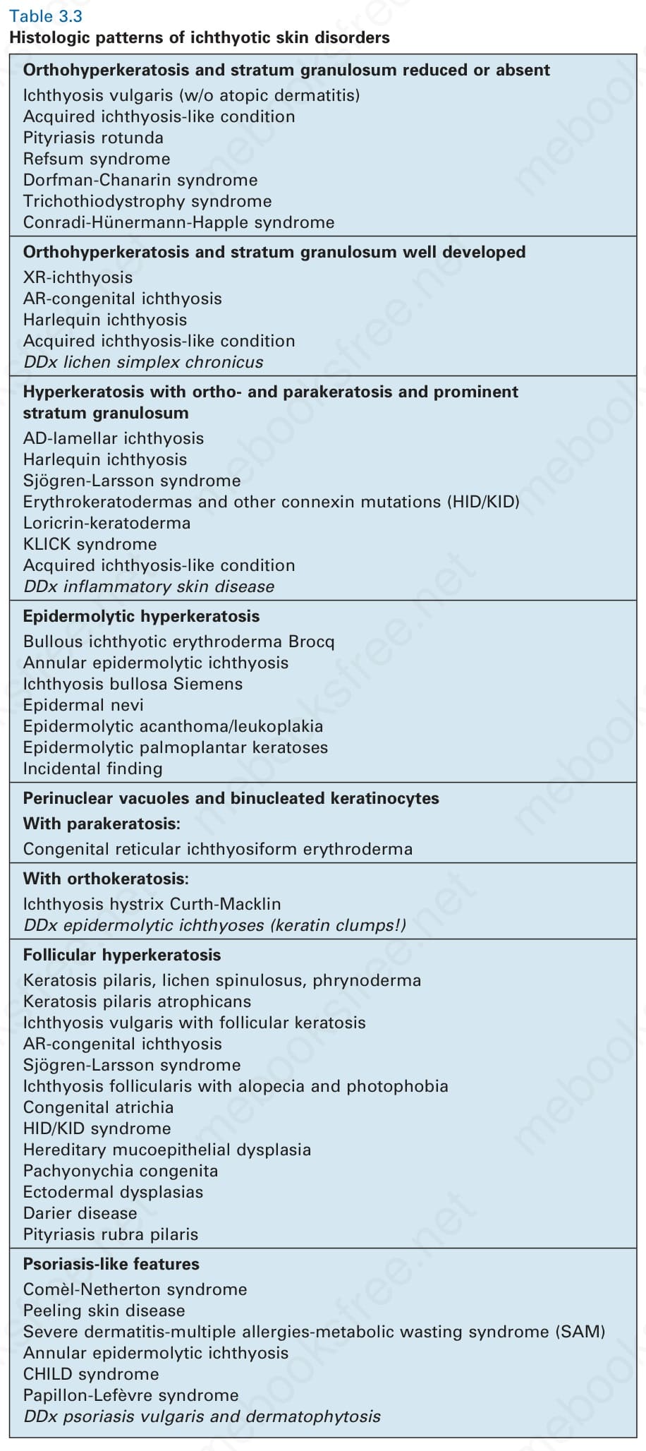
Table 3.3 Histologic patterns of ichthyotic skin disorders