Superficial epidermolytic ichthyosis
Superficial epidermolytic ichthyosis
Clinical features Superficial epidermolytic ichthyosis (SEI), formerly termed ichthyosis bullosa Siemens, is a keratinopathic ichthyosis, which is milder than EI. It presents at birth with blistering subsequently replaced by dark lichenified hyperkeratosis of the limbs, predominantly affecting the flexures and shins (Fig. 3.32).1,2 The skin remains fragile and blisters on mild trauma, giving rise to characteristic superficial peeling with a molting-like appearance (Mauserung phenomenon) (Fig. 3.33).2,3 Symptoms usually improve with age. Erythroderma is typically absent. Rarely, pustulation and hypertrichosis may be additional features.3,4 There is considerable clinical overlap between SEI and EI, and their distinction can best be achieved by molecular genetic analysis.4–10
66 Disorders of keratinization
Histologically and by electron microscopy, the features are indistinguishable from EI except that they are milder and the vacuolation of the keratinocytes and cytoplasmic inclusions are restricted to the more superficial spinous and granular cell layers. Subcorneal separation may be evident.11
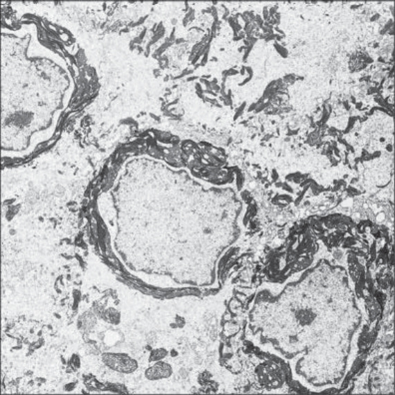
Fig. 3.30 Epidermolytic ichthyosis: striking perinuclear keratin clumping is evident. By courtesy of R.A.J. Eady, MD, Institute of Dermatology, London, UK.
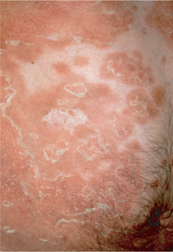
Fig. 3.31 Annular epidermolytic ichthyosis: migrating, polycyclic, gray hyperkeratotic plaques with a peripheral erythematous border. By courtesy of H. Traupe, MD, Münster, Germany.
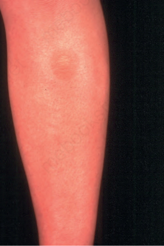
Fig. 3.32 Superficial epidermolytic ichthyosis: flexural hyperkeratosis with early blister formation. By courtesy of W.A.D. Griffiths, MD, Institute of Dermatology, London, UK.
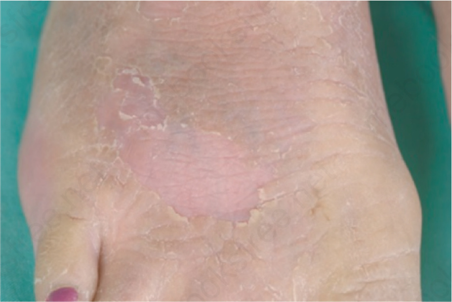
Fig. 3.33 Superficial epidermolytic ichthyosis: dark lichenified hyperkeratosis and characteristic superficial peeling with a molting-like appearance (Mauserung phenomenon). By courtesy of H. Traupe, MD, Münster, Germany.