Diagnosis of lymphomas
Diagnosis of lymphomas
The diagnosis and subclassification of lymphomas has transformed dramatically in the past three decades. Prior to this, the classifications in general use were based purely on the morphological features of the neoplastic lymphocytes.1,2 However, modern classification systems also utilize all available immunophenotypic, genetic, and clinical information to group cases together for the purposes of treatment and prognostication.3–7 Immunohistochemical and molecular techniques therefore form an integral part of the diagnostic process, and are routinely employed in the assessment of suspect cutaneous lymphoproliferations in order to discriminate reactive from neoplastic processes and to subclassify the latter once identified. A battery of antibodies and molecular techniques are now available to the practicing pathologist.8–14
For cancer, NGS can detect mutations, deletions, insertions, and copy number alterations across as many genes as desired. The main clinical application of this approach in dermatopathology has been with melanoma (Fig. 2.31).39–41 More specific melanoma applications will be discussed in greater detail in Chapter 26. There are balances that must be maintained between increasing the number of genes interrogated and the escalating costs and complexity of doing so. Some insight into fusion genes detection can be gained by DNA sequencing, but RNA approaches are the preferred methods as they are more sensitive and robust.42 NGS is currently the preferred sequencing method to broadly interrogate circulating (tumor) DNA also termed liquid biopsies that may someday substitute in some situations or complement tumor tissue–based diagnostics.43,44 RNA sequencing can also be used to determine gene expression similar to other hybridization and quantitative sequencing approaches.45 Micro- and long- noncoding RNA can also be detected if desired. This technique can be so sensitive that RNA sequencing can be done from single cells, but there are currently no clinical applications specifically for this approach.46 NGS has also transformed our ability to interrogate germ line DNA for variants or mutations linked to disease. These NGS-based approaches to cancer and heritable diseases are becoming widespread in the clinic and with the rapidly declining cost of NGS and improving reliability of interpretative bioinformatics algorithms, the clinical applications
This section focuses specifically on applications of the polymerase chain reaction (PCR) to diagnostic hematopathology, the molecular technique in most common usage, for the detection of antigen receptor gene rearrangement. The relevant immunophenotypic and genetic features of specific lymphoma subtypes are detailed in Chapter 29. In addition, FISH-based techniques can also be used to demonstrate translocation associated primarily with B-cell lymphomas.
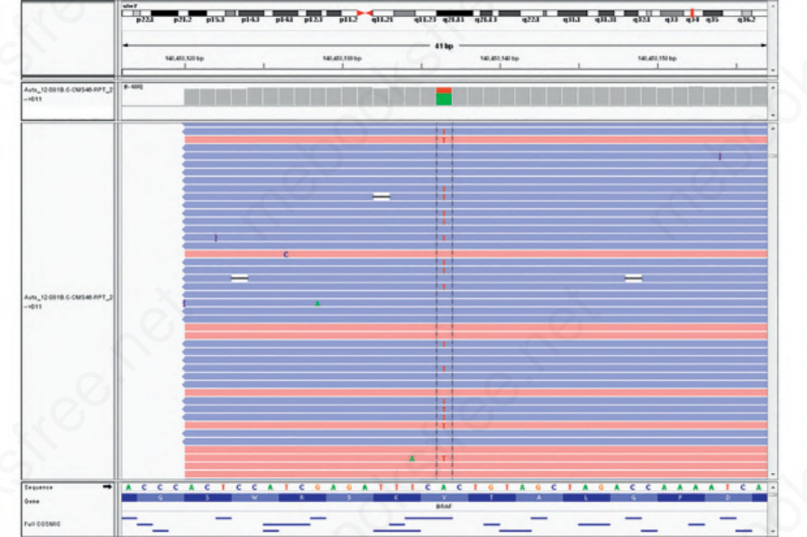
Fig. 2.31 Integrated genome viewer (IGV) depiction of a BRAF V600E mutation in melanoma. Note that the display shows individual DNA strands sequenced in both directions (forward and reverse strands, blue and red) by NGS sequencing. Thus the precise variant allele frequency can be determined.