Polymerase chain reaction (PCR)
Polymerase chain reaction (PCR)
In the diagnostic setting, PCR is used primarily to acquire sufficient DNA for analysis by sequencing or other methods, primarily to demonstrate a mutation or other genetic change or the presence of a specific gene or messenger RNA.
PCR is an extremely flexible technique and can be adapted to:
• detect mutations (base pair substitutions, insertions and deletions) in genes,
• demonstrate novel fusion transcripts (gene fusions),
• demonstrate clonality,
• demonstrate loss of heterozygosity (loss of one allele),
• detect DNA or RNA associated with infectious organisms,
• detect the levels of expression of messenger RNA. The ability to specifically amplify and detect any segment of DNA in the human genome has opened many diagnostic doors. In this technique, a pair of short sequences of DNA (called primers) that hybridize to two sequences of genomic DNA (or RNA reverse transcribed to DNA) are designed to amplify a specific region of DNA. Using a DNA polymerase that is stable at high temperatures, a series of annealing, extending, and melting/denaturing cycles amplifies the DNA between the two probes. This technique can be used on nucleic acids extracted from formalin-fixed, paraffin-embedded tissue, although probes must be designed to amplify shorter segments of DNA since the starting material has been cross-linked and fragmented from the formalin treatment. A variety of techniques based on PCR can be used to amplify DNA and then determine its sequence. Direct sequencing of genomic DNA allows detection of point mutations in cancer, such as BRAF or NRAS in melanoma.18 Generally, one can detect a mutation in 1 in 5 cells with this technique. More sensitive techniques such as pyrosequencing can reduce this to 1 in 10 or 20 cells by analysis for a precise mutation. Allele-specific PCR can be used to detect a known point mutation in as little as 1 in 50 or 100 cells. This technique has applications such as detecting KIT D816V mutation in mastocytosis in skin where the neoplastic cells may be sparse relative to the surrounding normal tissue.19 Digital droplet PCR can also provide high sensitivity and is focused on a single precise nucleotide change.20 Insertions and deletions in genes can also be detected, usually by Sanger sequencing.9 Real-time PCR allows indirect visualization of the desired PCR product (amplicon) through the amplification cycles and can provide rapid assessment of a positive or negative result.21
a gain. Gains with a high ratio that only affect portions of a chromosomal arm are termed amplifications. They arise from multiple independent events (chromosomal breakage and fusions) that accumulate under positive selection, typically because the genomic region present in the amplicon contained an oncogene, i.e., a gene that provided a growth advantage to the tumor cells with increased copies of the gene.
Reverse transcribing RNA to DNA can allow specific detection of fusion genes produced by chromosomal translocations such as seen in clear cell sarcoma or dermatofibrosarcoma protuberans.22,23 This technique is particularly valuable as there is no amplification product in the absence of tumor as the translocations are not seen in normal tissue or other tumors (see Fig. 2.29). Because the two genes involved in a translocation event can have breaks at a variety of introns (the noncoding region of DNA between the protein encoding exon segments), multiple primer pairs are often necessary to detect all of the possible translocation types. Also, since multiple genes can be involved, e.g., clear cell sarcoma can contain either an EWSR1-ATF1 or EWSR1-CREB1 fusion, additional primer sets will be required for detection of these as well (see Fig. 17.18C).24,25 In hematopoietic malignancies, detection of fusion transcripts can be used to detect minimal residual disease in the peripheral blood or marrow to measure tumor DNA as a surrogate of tumor load to assess response to therapy or allow early detection of recurrence. This approach may be applied to solid tumors in the future.
49 Molecular techniques
Primer Primer
Primer Primer
ATF1
EWSR1
EWSR1 / ATF1 Fusion
PCR can be used to detect normal genes as well. An instance of this in dermatopathology was the attempt to detect melanocyte-specific RNA (reverse transcribed to DNA) in sentinel lymph nodes that might have been missed by histology and immunohistochemical screening.26–28 Ultimately, this technique was not valuable, at least in part because of the presence of nodal nevi that would also be detected by this technique. While widely used in the research arena, other diagnostic approaches based on detection of gene expression will likely evolve with time.
Amplification No amplification
1
2
Type 2 EWSR1-ATF1 fusion amplicon
It is often advantageous to have multiple methodologies for detecting various molecular defects, as they are used in different situations and provide slightly different information. Fig. 2.30 depicts this for the translocation present in clear cell sarcoma.
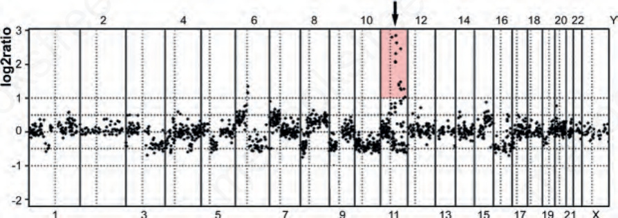
Fig. 2.28 DNA copy number changes as detected by array comparative genomic hybridization of an acral melanoma: the graph shows the log2 of the ratio of the fluorescence intensity ratios of tumor to reference DNA plotted according to their genomic position on the x-axis. The numbers at the top and at the bottom indicate the chromosomes. A log2 ratio of zero corresponds to normal copy number. As can be seen, multiple contiguous chromosomal regions showed losses and gains. The arrow corresponds to an amplification of chromosome 11q13 interval containing the gene that encodes cyclin D1.
Fig. 2.29 Use of RT-PCR to detect fusion transcripts: this technique uses reverse transcription to convert RNA to cDNA that can then be amplified by PCR. This step is necessary as the breakpoints in the usually large intronic regions of genomic DNA within a gene are essentially random, making it extremely difficult to amplify such large regions to identify the breakpoints using genomic DNA as the template. When the gene is transcribed to RNA, the introns are removed during splicing and introns are directly juxtaposed (Fig. 2.22C) allowing more ready detection of the novel juxtaposition of exons from two different genes. When primers are designed for the exons of each of the two genes involved in a translocation, amplification only occurs of the cDNA of the fusion transcript as these introns would not be adjacent in normal tissue. This product will have a specific size and can be detected on a gel, but direct DNA sequencing or other methods should be used to confirm its identity. Amplification of normal housekeeping gene transcripts are used to ensure the quality of the cDNA.
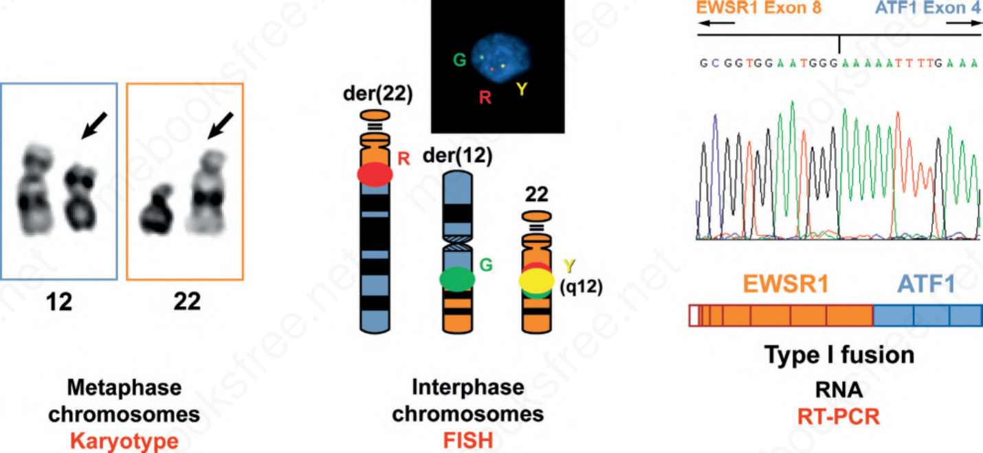
Fig. 2.30 Multiple modalities for detection of recurrent translocations. Traditional karyotypes use metaphase chromosomes spreads to detect translocations and other structural genetic aberrations using banding (staining) techniques. FISH uses less condensed interphase chromosomes to detect rearrangements or amplifications. RT-PCR can detect the precise exons involved in a fusion RNA transcript. Each is a valid method for demonstrating chromosomal translocations, but each has applicability to different sample types and provides different information.