Electron microscopy
Electron microscopy
Electron microscopy is much less utilized than in the past. Immunohistochemical approaches are preferred in those instances where they are a reasonable substitute.
Transmission electron microscopy offers much better resolution than light microscopy.1 To optimize this, tissue has to be embedded in extremely rigid material to allow ultrathin sectioning at 80 nm. In most circumstances, hydrophobic epoxy resins are preferred. When a specimen requires ultrastructural examination, the portion to be examined must be treated in a suitable fixative immediately. The volume of the fixative should be 10 times the sample size. The final specimen is cubed to 1 mm portions.1 Fixation is affected by:
• pH,
• osmolarity,
• ionic composition of buffer,
• fixative concentration,
• temperature,
• duration of fixation. Primary fixation in an aldehyde, usually glutaraldehyde, and secondary fixation in osmium tetroxide are standard procedures. Advances in immunohistochemistry have decreased the dependence on electron microscopy for ultrastructural confirmation of cell lineage. Notwithstanding, dermatologic ultrastructural investigations are important in the diagnosis of:
• undifferentiated tumors,
• immunobullous disease,
• cerebral autosomal dominant arteriopathy with subcortical infarcts and leukoencephalopathy (CADASIL),
• amyloidosis,
• metabolic storage diseases.2–7
Intercellular junctions, Weibel-Palade bodies, melanosomes, and premelanosomes may help in the diagnosis of carcinomas, endothelial tumors, and melanocytic tumors, respectively.3 In CADASIL, extracellular, electron-dense granular material is present in an indentation in vascular smooth muscle cells.5,6 Amyloid is identifiable as randomly arranged, extracellular, nonbranching fibrils of indeterminate length and 7–10 nm diameter.7 Transmission electron microscopy remains a valuable tool in the ongoing evaluation of the structure of normal and pathological human cell and tissue components and infective agents.8–10 Technological advancements have enabled electronic capture of ultrastructural images (Fig. 2.14).
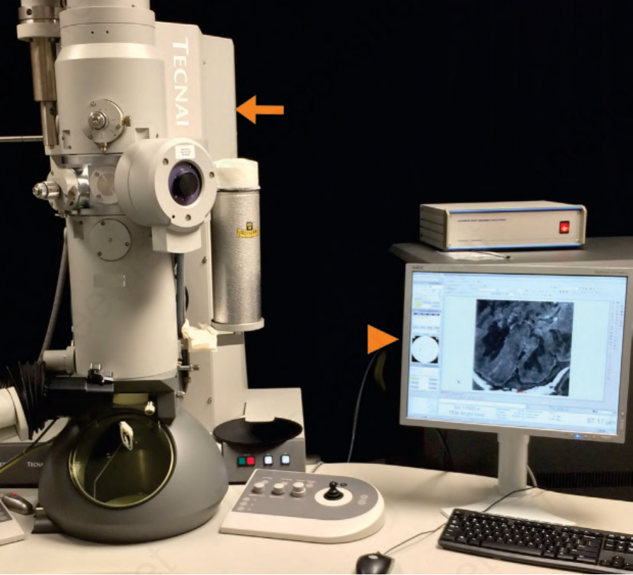
Fig. 2.14 Transmission electron microscope (arrow) with electronic image capture (arrowhead).