疾病定義與分類
- 巨細胞血管母細胞瘤 (giant cell angioblastoma) 是否為真正的血管腫瘤目前仍不明確,在此僅作簡要描述,待更多文獻釐清其分化譜系 (line of differentiation)。
- 目前僅有極少數病例被報導,多數出現於兒童的下肢與骨骼。腫瘤體積大,且呈進行性生長。曾有單一成人病例的報導。
組織病理特徵 (Histopathology)
- 組織學顯示以結節 (nodules) 形式分布、環繞血管腔道 (vascular channels) 的類組織球細胞 (histiocyte-like cells) 聚集。
- 約 30% 的病例可見明顯的嗜中性球發炎浸潤 (neutrophilic inflammatory infiltrate)。壞死 (necrosis) 與血管侵犯 (vascular invasion) 罕見。
免疫組化與特殊染色 (Immunohistochemistry & Special Stains)
- 免疫組織化學上,腫瘤細胞對 CD31、ERG、FLI1 及 keratin AE1/AE3 呈陽性;對 CD34、epithelial membrane antigen (EMA) 及其他 keratins(包括 MNF116)呈陰性。
- 可見局部 SMA 陽性,而 S100 與 desmin 呈陰性染色。
- 腫瘤細胞對 FOSB 呈現強烈的核陽性。此後者標記在一部分 epithelioid hemangiomas 亦呈陽性,且罕見於 nodular 與 proliferative fasciitis 的病例中,但不出現於其他可能與 pseudomyogenic hemangioendothelioma 混淆的血管腫瘤。
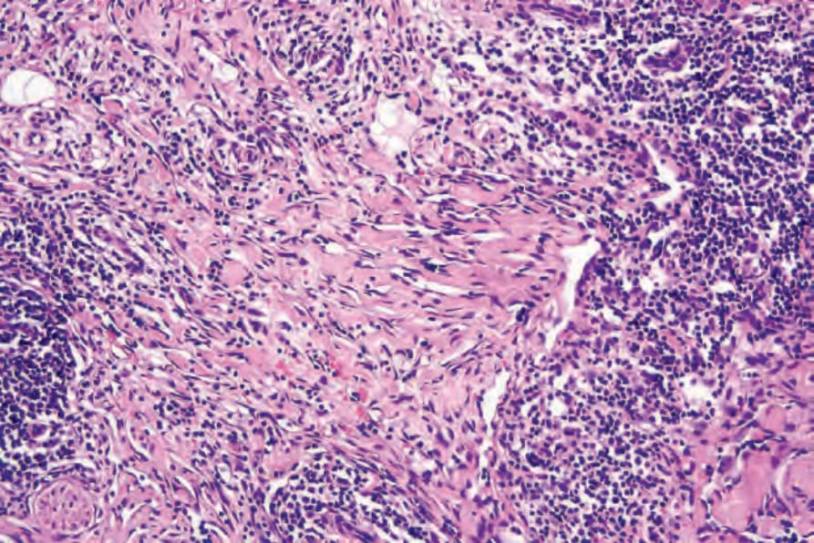
圖 35-557:複合性血管內皮瘤 (composite hemangioendothelioma):可見明顯的膠原蛋白剝離 (dissection of collagen),類似血管肉瘤 (angiosarcoma)。
Fig. 35.557 Composite hemangioendothelioma: there is marked dissection of collagen suggestive of angiosarcoma.
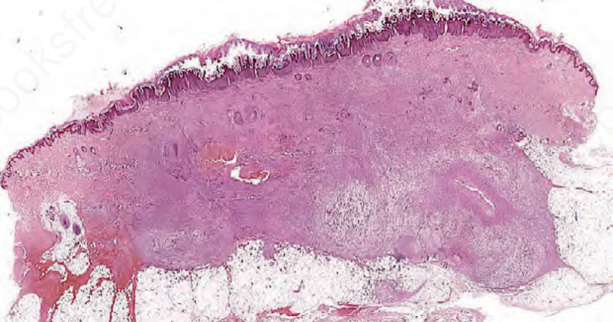
圖 35-558:假肌源性血管內皮瘤 (pseudomyogenic hemangioendothelioma):浸潤性腫瘤明顯侵犯真皮與皮下組織。
Fig. 35.558 Pseudomyogenic hemangioendothelioma: prominent involvement of the dermis and subcutaneous tissue by an infiltrative tumor.
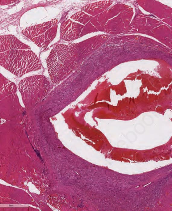
圖 35-559:假肌源性血管內皮瘤 (pseudomyogenic hemangioendothelioma):與前圖為同一病人。多病灶性腫瘤亦侵犯骨骼肌 (skeletal muscle)。
Fig. 35.559 Pseudomyogenic hemangioendothelioma: same patient as previous figure. Multifocal tumor also involving skeletal muscle.
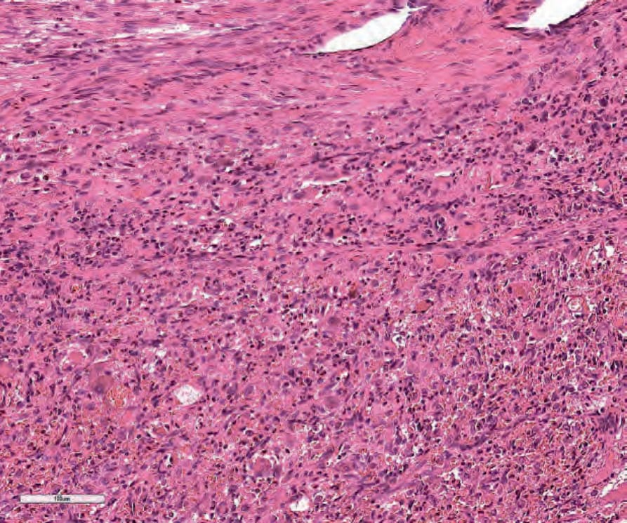
圖 35-560:假肌源性血管內皮瘤 (pseudomyogenic hemangioendothelioma):圓形的假橫紋肌母細胞 (pseudorhabdomyoblast) 與梭形細胞,無提示血管分化的特徵。
Fig. 35.560 Pseudomyogenic hemangioendothelioma: round pseudorhabdomyblast and spindle-shaped cells with no features suggestive of vascular differentiation.