Giant cell angioblastoma
Giant cell angioblastoma
It is unclear whether this is a true vascular tumor but it is briefly described here until further reports clarify the line of differentiation. A very small number of cases have been reported, most presenting in the lower limbs of children and in bone.1–3 Tumors are large and present with progressive growth.1,2 A single case has been described in an adult.4
Histology shows aggregates of histiocyte-like cells in nodules distributed around vascular channels.
1855 Malignant vascular tumors
A prominent neutrophilic inflammatory infiltrate identified in about 30% of cases. Necrosis and vascular invasion are rare.
Immunohistochemically, tumor cells are positive for CD31, ERG, FLI1 and keratin AE1/AE3, and negative for CD34, epithelial membrane antigen (EMA), and other keratins including MNF116.1 Focal positivity for SMA may be seen and there is negative staining for S100 and desmin. Tumor cells display strong nuclear positivity for FOSB.16–19 The later marker is also positive in a percentage of epithelioid hemangiomas and rarely in cases of nodular and proliferative fasciitis but not in other vascular tumors that may be confused with pseudomyogenic hemangioendothelioma.
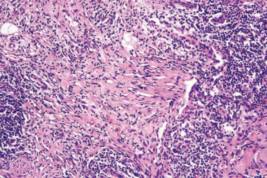
Fig. 35.557 Composite hemangioendothelioma: there is marked dissection of collagen suggestive of angiosarcoma.
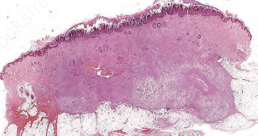
Fig. 35.558 Pseudomyogenic hemangioendothelioma: prominent involvement of the dermis and subcutaneous tissue by an infiltrative tumor.
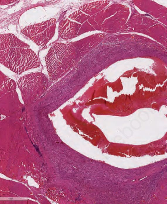
Fig. 35.559 Pseudomyogenic hemangioendothelioma: same patient as previous figure. Multifocal tumor also involving skeletal muscle.
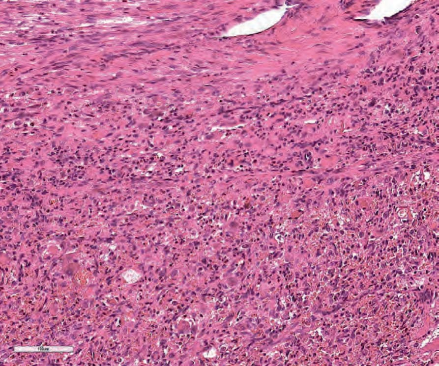
Fig. 35.560 Pseudomyogenic hemangioendothelioma: round pseudorhabdomyblast and spindle-shaped cells with no features suggestive of vascular differentiation.