頸部胸腺囊腫 (Cervical Thymic Cyst)
臨床特徵 (Clinical Features)
頸部胸腺囊腫 (cervical thymic cyst) 大多侵犯兒童,沿著從下頜角 (angle of the mandible) 至胸骨柄 (manubrium sternae) 的連線、出現於頸前三角 (anterior triangle)。亦可見表現於較深部組織者。此類病灶占先天性頸部腫塊 (congenital neck masses) 的百分之零點三至百分之一。病灶僅在極少數情況下出現於成人。頸部左側受侵犯者占百分之六十八,右側占百分之二十五,中線占百分之七。延伸進入縱膈腔 (mediastinum) 者甚為常見。囊腫可能在 Valsalva 動作 (Valsalva maneuver) 時增大。
致病機轉與組織學特徵 (Pathogenesis and Histologic Features)
此囊腫由胸腺咽管 (thymopharyngeal duct) 的殘餘構造發育而成;在胸腺前驅組織下降進入縱膈腔時,這些殘餘構造持續存在。
囊腫為單房 (unilocular) 或多房 (multilocular),並具有腔內乳頭狀突起 (intraluminal papillary projections)(Fig. 34.33)。其襯裡與正常輸卵管 (fallopian tube) 相似,由立方形至柱狀的纖毛上皮 (cuboidal to columnar ciliated epithelium) 構成,並常見偽複層 (pseudostratified) 病灶(Fig. 34.34)。偶亦可見散在的暗細胞 (intercalated dark cells)。鱗狀化生 (squamous metaplasia) 常為其特徵之一。分泌黏液之細胞 (mucin-secreting cells) 曾於極例外情況下被描述,並有一兩篇報告記載類頂泌汗腺 (apocrine-like) 特徵。上皮深部為血管化良好的平行膠原束 (well-vascularized parallel bundles of collagen),但平滑肌 (smooth muscle) 並非其特徵。
在超微結構上,纖毛具有特徵性的形態,包含一對中央微管 (central pair of microtubules)、九對放射狀排列的微管 (nine radially orientated pairs of microtubules)、基體 (basal bodies),以及橫紋小根 (cross-striated rootlets)。有時亦可見微絨毛 (microvilli)。
免疫組化與特殊染色 (Immunohistochemistry & Special Stains)
襯裡細胞表現 keratin 與上皮膜抗原 (epithelial membrane antigen, EMA),但不表現 CEA、desmin 與平滑肌肌動蛋白 (smooth muscle actin, SMA)。曾有一例出現於男性面頰、並顯示 S100 protein 與 SMA 陽性之肌上皮層 (myoepithelial layer),該例較適合歸類為伴有纖毛化生之汗腺汗囊瘤 (sweat gland hidrocystoma with cilia metaplasia)。曾有一例記載 desmin 表現侷限於纖毛細胞的頂端面 (apical aspect)。雌激素 (estrogen) 與黃體素受體 (progesterone receptors) 可能為陽性,且偶可見 S100 protein。在發生於女性的病灶中亦曾報告 PAX8 與 WT-1 陽性,支持這些病灶具有 Müllerian 分化 (Müllerian differentiation)。單一病例報告亦記載 amylase 與 dynein 的表現。
鑑別診斷 (Differential Diagnosis)
纖毛上皮細胞 (ciliated epithelial cells) 亦可見於支氣管源性囊腫 (bronchogenic cyst)、甲狀舌管囊腫 (thyroglossal duct cyst)、鰓裂囊腫 (branchial cyst) 與胸腺囊腫 (thymic cyst)。它們也可能出現於成熟囊性畸胎瘤 (mature cystic teratoma)。
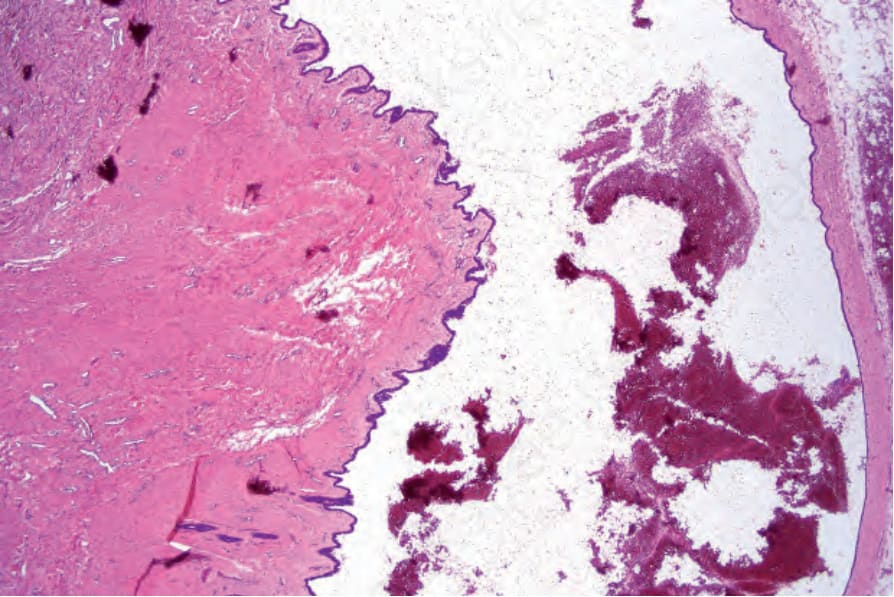
圖 34-33:皮膚纖毛囊腫 (cutaneous ciliated cyst):注意視野左下方的乳頭狀突起 (papillary projections)。
Fig. 34.33 Cutaneous ciliated cyst: note the papillary projections in the lower left of the field.
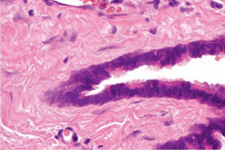
圖 34-34:皮膚纖毛囊腫 (cutaneous ciliated cyst):囊壁由高柱狀細胞 (tall columnar cells) 襯覆。視野中央可見纖毛 (cilia)。
Fig. 34.34 Cutaneous ciliated cyst: the wall is lined by tall columnar cells. Cilia are evident in the center of the field.
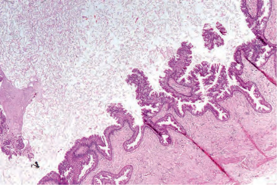
圖 34-35:正中縫囊腫 (median raphe cyst):低倍視野,顯示由柱狀上皮 (columnar epithelium) 覆蓋的乳頭狀突起 (papillary processes)。
Fig. 34.35 Median raphe cyst: low-power view showing papillary processes covered by columnar epithelium.