Cervical thymic cyst
Cervical thymic cyst
Clinical features Cervical thymic cysts mostly affect children and present in the anterior triangle along a line from the angle of the mandible to the manubrium sternae.1–6 Presentation in deeper tissues may also be seen. Lesions account for 0.3% to 1% of congenital neck masses.7 Lesions only exceptionally present in adults.8 The left side of the neck is affected in 68% of cases, the right side in 25% of cases, and the midline in 7% of cases.3 Extension into the mediastinum is common.5 The cyst may enlarge on Valsalva maneuver.
Pathogenesis and histologic features The cyst develops from remnants of the thymopharyngeal duct which persist as the thymic precursor descends into the mediastinum.3,4
The cyst is unilocular or multilocular and has intraluminal papillary projections (Fig. 34.33). The lining, which is similar to that of normal fallopian tube, consists of cuboidal to columnar ciliated epithelium with frequent pseudostratified foci (Fig. 34.34).6 Intercalated dark cells are also occasionally evident.4 Squamous metaplasia is often a feature. Mucin-secreting cells have very exceptionally been described, and there are one or two reports of apocrine-like features.2,8,11 Deep to the epithelium lie well-vascularized parallel bundles of collagen, but smooth muscle is not a feature.1
Ultrastructurally, the cilia have characteristic morphology with a central pair of microtubules, nine radially orientated pairs of microtubules,
1693 Glandular cysts
basal bodies, and cross-striated rootlets.7,8,13 Microvilli are sometimes evident.8,14
The lining cells express keratin and epithelial membrane antigen (EMA) but not CEA or desmin and smooth muscle actin (SMA).8–10,14,15 One case, which presented on the cheek of a male and demonstrated an S100 protein and SMA positive myoepithelial layer, would be better classified as a sweat gland hidrocystoma with cilia metaplasia.21 Desmin expression restricted to the apical aspect of the ciliated cells has been documented in one case.8 Estrogen and progesterone receptors may be positive and S100 protein is occasionally present.8,12,14 Positivity for PAX8 and WT-1 has also been reported in lesions arising in females supporting Müllerian differentiation in these lesions. 25–27 Single case reports also document expression of amylase and dynein.3,10
Differential diagnosis Ciliated epithelial cells are also seen in bronchogenic, thyroglossal duct, branchial, and thymic cysts. They may also be present in mature cystic teratoma.
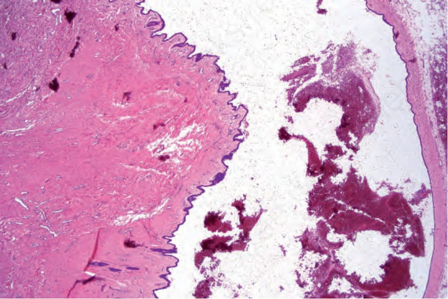
Fig. 34.33 Cutaneous ciliated cyst: note the papillary projections in the lower left of the field.
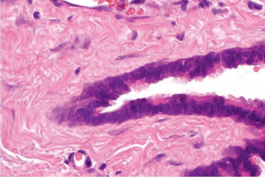
Fig. 34.34 Cutaneous ciliated cyst: the wall is lined by tall columnar cells. Cilia are evident in the center of the field.
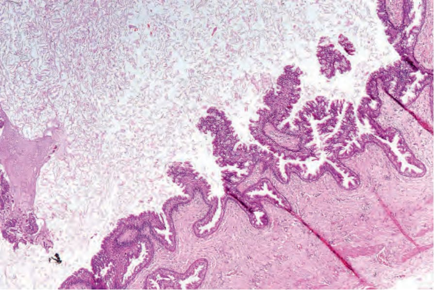
Fig. 34.35 Median raphe cyst: low-power view showing papillary processes covered by columnar epithelium.