臨床特徵 (Clinical Features)
- 結膜藍痣 (conjunctival blue nevus) 與細胞性藍痣 (cellular blue nevus) 為罕見病灶,源自胚胎遷移過程中未抵達上皮表面的神經嵴細胞 (neural crest cells)。
- 臨床上病灶界線分明,外觀呈棕色或黑色。病灶可為單灶性 (unifocal) 或多灶性 (multifocal),可發生於結膜任何部位。
- 目前僅有一例自 blue nevus 發生惡性黑色素瘤 (malignant melanoma) 的報告。
- (此特徵)並不代表惡性。在其他方面屬於典型的結膜痣 (conjunctival nevi) 中,可見兩種不同的痣細胞 (nevus cells):氣球細胞 (balloon cells) 與梭形細胞 (spindle cells)。氣球細胞痣 (balloon cell nevi) 極少被報告。偶爾會遇到結膜複合痣 (conjunctival combined nevi)。
組織病理特徵 (Histopathology)
- 結膜 blue nevus 由位於固有質 (substantia propria) 內、色素均勻分布的梭形細胞 (spindle-shaped cells) 構成,並可見樹突狀黑色素細胞 (dendritic melanocytes)。
- 細胞性藍痣 (cellular blue nevus) 具有由均勻梭形細胞組成、輕度色素化的結節,周圍環繞著重度色素化的樹突狀黑色素細胞 (dendritic melanocytes)。
- 當 blue nevus 的成分與結膜的一般後天性痣 (common acquired nevus) 或先天性痣 (congenital nevus) 同時存在時,則稱為複合痣 (combined nevus)(Fig. 27.31)。
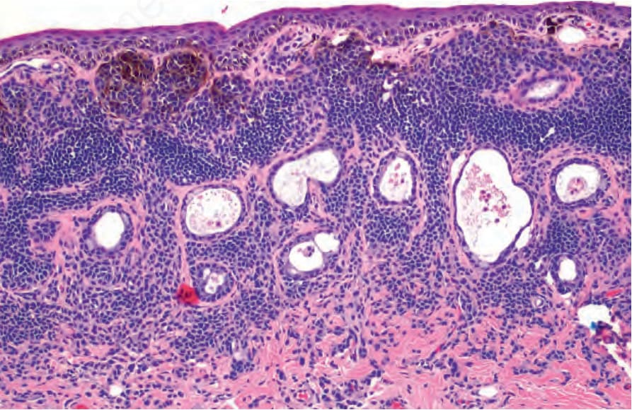
圖 27-30:上皮下痣 (subepithelial nevus):痣細胞 (nevus cells) 存在於固有質 (substantia propria) 內,但無交界處巢狀結構 (junctional nests)。病灶內可見上皮性囊腫 (epithelial cysts)。請注意表淺處痣細胞的黑色素 (melanin) 色素化程度較朝向基底處的痣細胞為高。
Fig. 27.30 Subepithelial nevus: nevus cells are present in the substantia propria without junctional nests. Epithelial cysts are present within the lesion. Note how there is increased melanin pigmentation of the superficial nevus cells compared to those toward the base.
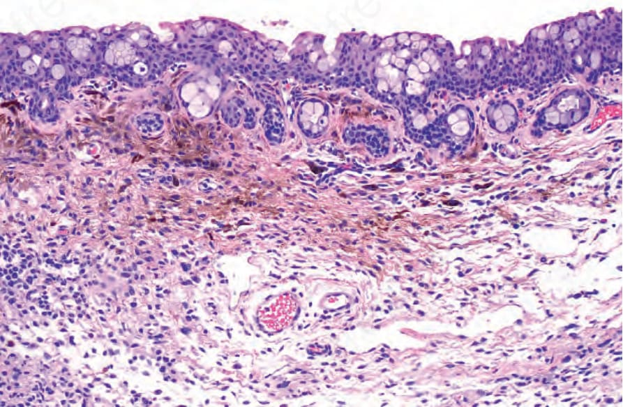
圖 27-31:複合痣 (combined nevus):組織學顯示一般痣細胞 (common nevus cells) 的巢狀結構,以及色素化程度較高的梭形與樹突狀細胞 (spindle and dendritic cells)。
Fig. 27.31 Combined nevus: histology shows nests of common nevus cells and more pigmented spindle and dendritic cells.