Blue nevus
Blue nevus
Clinical features Conjunctival blue and cellular blue nevi are rare lesions that arise from neural crest cells that do not reach the epithelial surface during embryonic migration. Clinically, the lesion is sharply demarcated and appears brown or black.1 Lesions may be unifocal or multifocal and occur at any site of the conjunctiva.2 There is only one reported case of malignant melanoma arising from blue nevus.3
not indicative of malignancy. Two distinct types of nevus cells, balloon cells and spindle cells, can be seen in otherwise typical conjunctival nevi. Balloon cell nevi have rarely been reported.3 Occasionally, conjunctival combined nevi are encountered.4
Histologic features Conjunctival blue nevus is composed of uniformly pigmented spindle-shaped cells in the substantia propria. Dendritic melanocytes are also present. Cellular blue nevus has lightly pigmented nodules of uniform spindle cells surrounded by heavily pigmented dendritic melanocytes.3 When elements of blue nevus are present with a common acquired or congenital nevus of the conjunctiva, it is termed combined nevus (Fig. 27.31).4
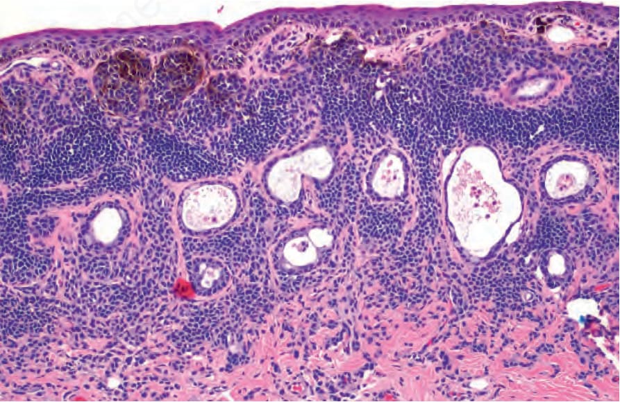
Fig. 27.30 Subepithelial nevus: nevus cells are present in the substantia propria without junctional nests. Epithelial cysts are present within the lesion. Note how there is increased melanin pigmentation of the superficial nevus cells compared to those toward the base.
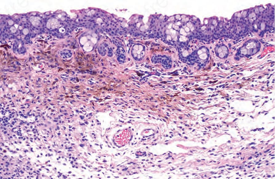
Fig. 27.31 Combined nevus: histology shows nests of common nevus cells and more pigmented spindle and dendritic cells.