上皮性囊腫 (Epithelial Cysts)
上皮性囊腫 (epithelial cysts)
臨床特徵 (Clinical Features)
- 結膜囊腫 (conjunctival cysts) 為常見病灶,可為先天性,但最常見為後天獲得性。其可自發發生,亦可於手術或非手術性外傷後出現。後天獲得性囊腫多為表面上皮的植入性 (implantation,即上皮包涵性 epithelial inclusion) 囊腫。源自副淚腺 (accessory lacrimal glands) 的導管囊腫 (ductal cysts) 亦相當常見。囊腫外觀平滑而半透明,內含清澈液體 (Fig. 27.15)。
組織病理特徵 (Histopathology)
-
角化性斑塊 (keratotic plaque) 由具角化 (keratinization) 與角化不全 (parakeratosis) 的棘層肥厚上皮 (acanthotic epithelium) 構成。光化性角化病 (actinic keratosis) 與之相似,且常發生於慢性發炎的瞼裂斑 (pinguecula) 或翼狀贅片 (pterygium) 之上。其細胞學異型性 (cytological atypia) 程度不一,可由極輕微至重度 (Fig. 27.17)。
-
上皮包涵性囊腫 (epithelial inclusion cysts) 內襯結膜上皮 (conjunctival epithelium),內含清澈液體。導管囊腫 (ductal cysts) 內襯雙層細胞,內含過碘酸-希夫染色 (periodic acid-Schiff, PAS) 陽性物質 (Fig. 27.16)。
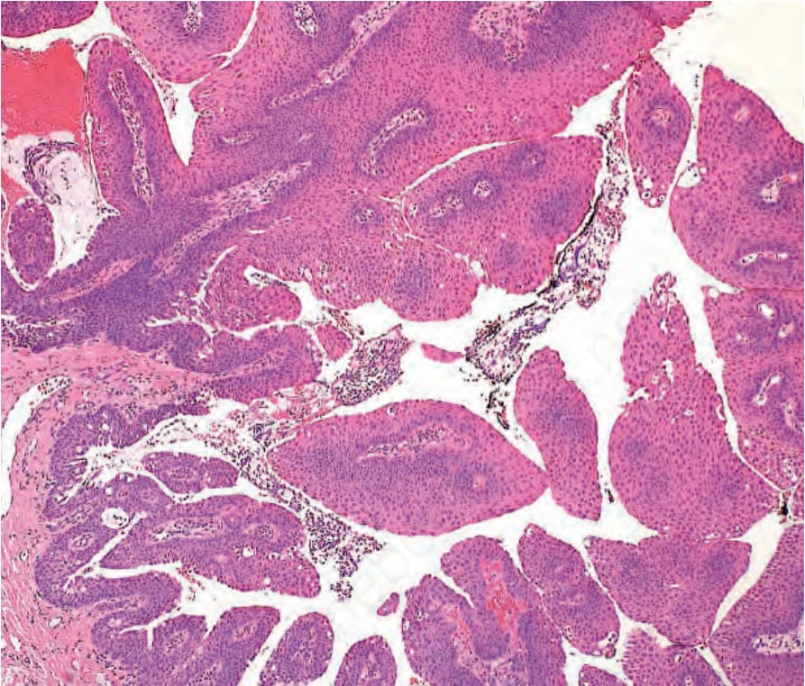
圖 27-12:鱗狀乳頭瘤 (squamous papilloma):組織學顯示由非角化鱗狀上皮 (nonkeratinized squamous epithelium) 構成的指狀突起,環繞中央的纖維血管軸心 (fibrovascular cores)。
Fig. 27.12 Squamous papilloma: histology shows fronds of nonkeratinized squamous epithelium surrounding central fibrovascular cores.
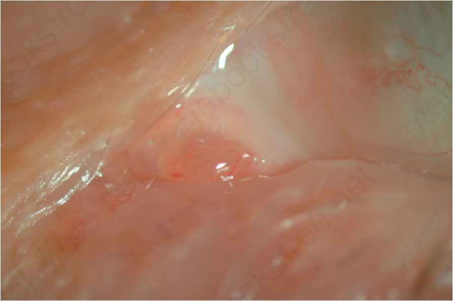
圖 27-13:嗜酸細胞瘤 (oncocytoma):位於淚阜 (caruncle) 的上皮下、肉質橙粉紅色病灶。
Fig. 27.13 Oncocytoma: subepithelial, fleshy orange-pink lesion in the caruncle.
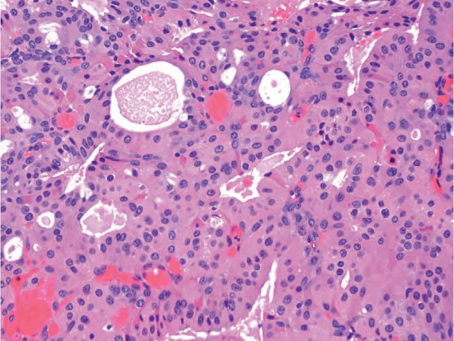
圖 27-14:嗜酸細胞瘤 (oncocytoma):淚阜 (caruncle) 固有層 (substantia propria) 內病灶的組織學,顯示具嗜酸性細胞質 (eosinophilic cytoplasm) 的大型細胞排列成腺體結構。
Fig. 27.14 Oncocytoma: histology of a lesion in the substantia propria of the caruncle shows large cells with eosinophilic cytoplasm arranged in glandular structures.
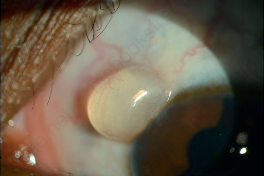
圖 27-15:上皮性囊腫 (epithelial cyst):鄰近角膜緣 (limbus) 之囊腫的臨床外觀。
Fig. 27.15 Epithelial cyst: clinical appearance of a cyst adjacent to the limbus.
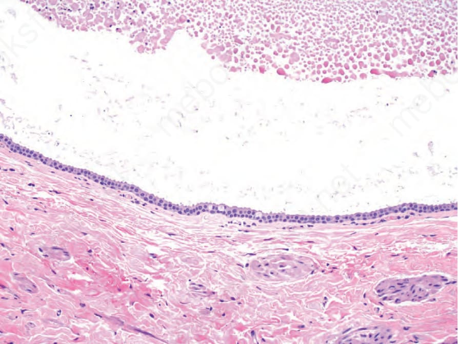
圖 27-16:上皮性囊腫 (epithelial cyst):組織學顯示囊腔內充滿上皮碎屑 (epithelial debris),內襯兩層立方上皮 (cuboidal epithelium),並偶見杯狀細胞 (goblet cells)。
Fig. 27.16 Epithelial cyst: histology shows a cystic space filled with epithelial debris and lined by two layers of cuboidal epithelium with rare goblet cells.
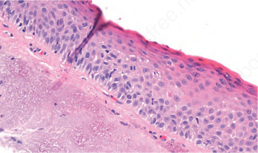
圖 27-17:光化性角化病 (actinic keratosis):組織學顯示棘層肥厚上皮 (acanthotic epithelium) 伴表面角化 (surface keratinization),及輕度細胞學異型性 (mild cytological atypia) 與有絲分裂活性增加。固有層 (substantia propria) 內可見光化性彈性纖維變性 (actinic elastotic degeneration)。
Fig. 27.17 Actinic keratosis: histology shows acanthotic epithelium with surface keratinization and mild cytological atypia with increased mitotic activity. Actinic elastotic degeneration is present in the substantia propria.