Epithelial cysts
Epithelial cysts
Clinical features Conjunctival cysts are common lesions that may be congenital or most often, acquired. They may occur spontaneously, or after surgical or nonsurgical
1369 Epithelial tumors
trauma.1,2 Acquired cysts are mostly implantation (epithelial inclusion) cysts of surface epithelium. Ductal cysts, which arise from accessory lacrimal glands, are also common. Cysts appear smooth and translucent, and contain clear fluid (Fig. 27.15).
Histologic features
Keratotic plaque is composed of acanthotic epithelium with keratinization and parakeratosis. Actinic keratosis is similar, and often occurs over a chronically inflamed pinguecula or pterygium.1–3 There is variable cytological atypia that ranges from minimal to severe (Fig. 27.17).1
Histologic features Epithelial inclusion cysts are lined by conjunctival epithelium and contain clear fluid. Ductal cysts are lined by a double layer and contain periodic acid-Schiff (PAS)-positive material (Fig. 27.16).
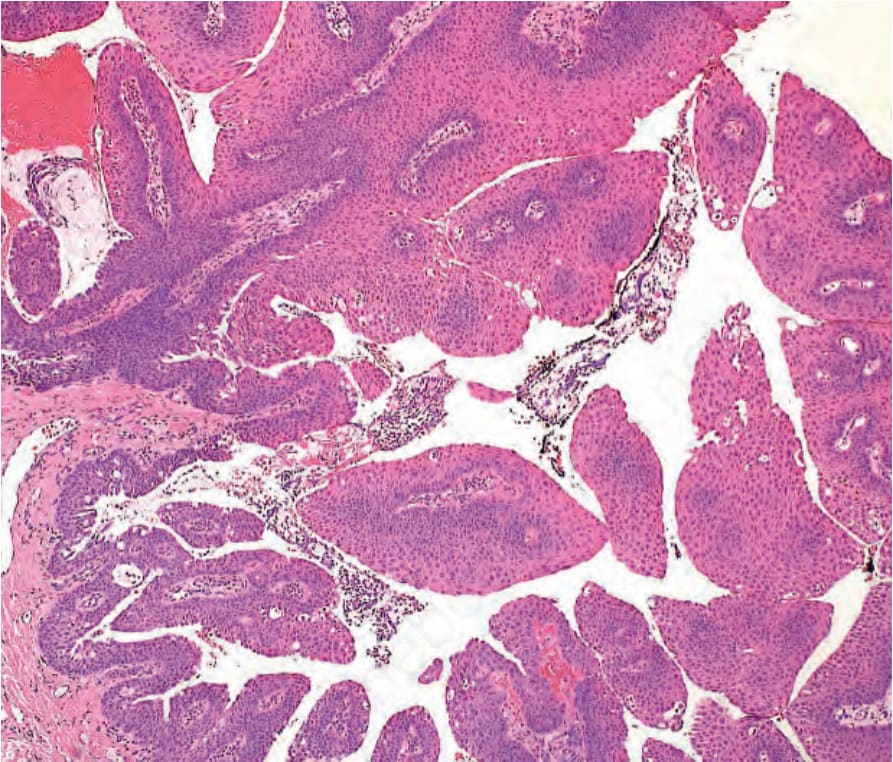
Fig. 27.12 Squamous papilloma: histology shows fronds of nonkeratinized squamous epithelium surrounding central fibrovascular cores.
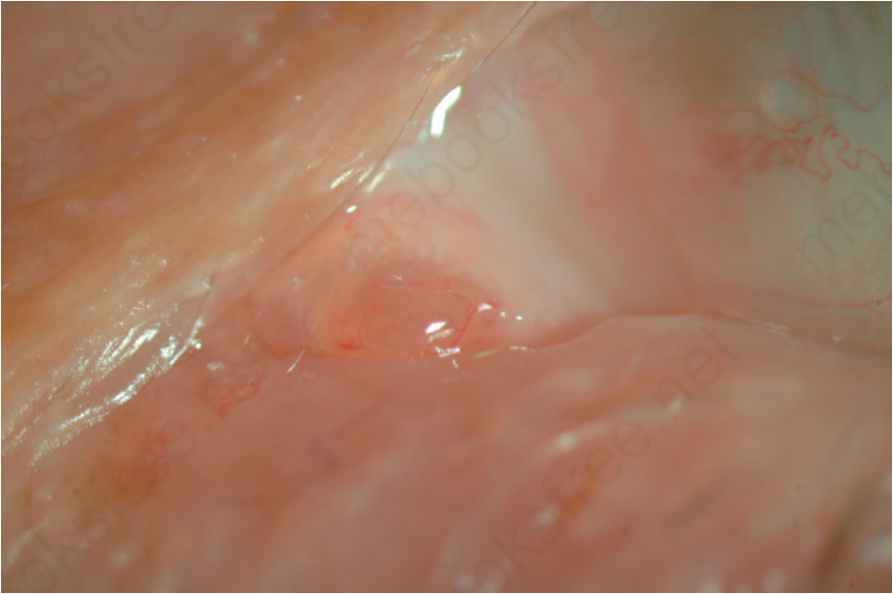
Fig. 27.13 Oncocytoma: subepithelial, fleshy orange-pink lesion in the caruncle.
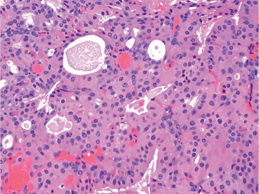
Fig. 27.14 Oncocytoma: histology of a lesion in the substantia propria of the caruncle shows large cells with eosinophilic cytoplasm arranged in glandular structures.
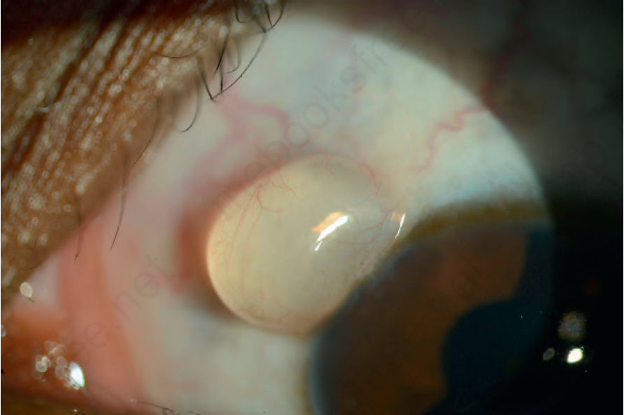
Fig. 27.15 Epithelial cyst: clinical appearance of a cyst adjacent to the limbus.
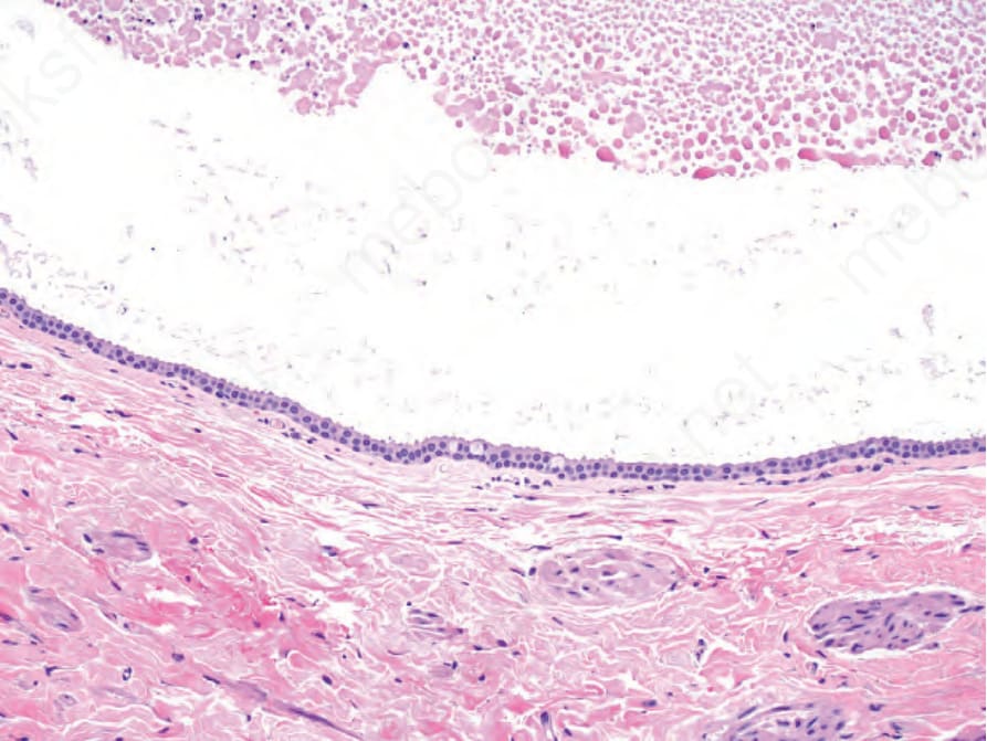
Fig. 27.16 Epithelial cyst: histology shows a cystic space filled with epithelial debris and lined by two layers of cuboidal epithelium with rare goblet cells.
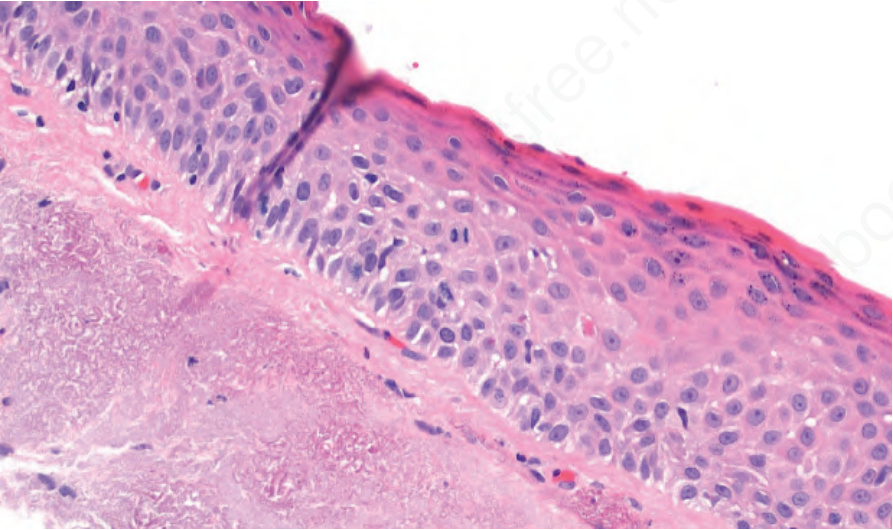
Fig. 27.17 Actinic keratosis: histology shows acanthotic epithelium with surface keratinization and mild cytological atypia with increased mitotic activity. Actinic elastotic degeneration is present in the substantia propria.