臨床特徵 (Clinical Features)
- 眼球表面皮樣瘤 (epibulbar dermoid)(Fig. 27.6)為一種先天性病灶,最常位於角膜緣 (limbus) 的下方顳側 (inferotemporally)。
- 病灶界線清楚 (well circumscribed),呈黃褐色 (tan-yellow),表面可能有細毛突出。
- 病灶可為單獨發生,或與 Goldenhar syndrome(眼–耳–脊椎發育不良 (oculoauriculovertebral dysplasia))相關。
- 患者應評估是否有耳前皮膚附屬器 (preauricular skin appendages)、聽力喪失 (hearing loss)、眼瞼缺損 (eyelid colobomas)、下頜骨發育不全 (mandibular hypoplasia) 及脊椎異常 (vertebral abnormalities)。
- Dermoid 可造成散光 (astigmatism) 與弱視 (amblyopia)。
組織病理特徵 (Histopathology)
- 複合型迷芽瘤 (complex choristomas) 可包含多種異位組織 (ectopic tissues) 的組合,包括含脂肪組織 (adipose tissue) 與皮膚附屬器 (dermal appendages) 的真皮組織、淚腺組織 (lacrimal gland tissue)、平滑肌 (smooth muscle)、軟骨 (cartilage) 及骨 (bone)(Figs 27.9 與 27.10)。
- Epibulbar dermoid 由緻密纖維組織 (dense fibrous tissue) 構成,其中含有皮膚附屬器,例如毛囊 (hair follicles)、皮脂腺 (sebaceous glands)、汗腺 (sweat glands),有時還有脂肪組織 (adipose tissue),並由非角化複層鱗狀上皮 (nonkeratinizing stratified squamous epithelium) 所襯覆(Fig. 27.7)。
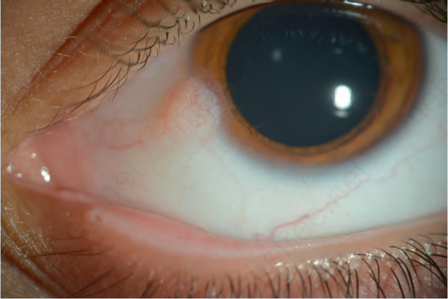
圖 27-6:角膜緣皮樣瘤 (limbal dermoid):臨床照片顯示位於角膜緣下鼻側 (inferonasally) 的隆起性黃白色病灶 (elevated, yellow-white lesion)。
Fig. 27.6 Limbal dermoid: clinical photograph showing an elevated, yellow-white lesion at the limbus inferonasally.
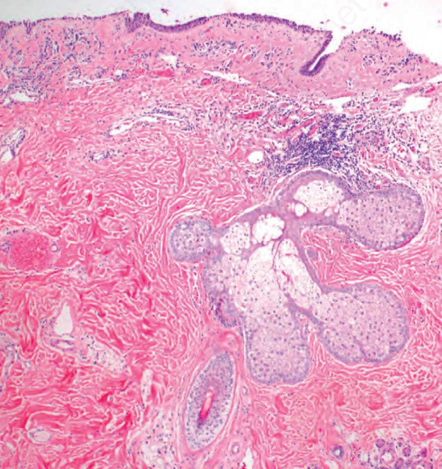
圖 27-7:角膜緣皮樣瘤 (limbal dermoid):組織學顯示真皮型膠原 (dermal type collagen) 與皮膚附屬器結構 (skin adnexal structures),包括毛囊 (hair follicle) 與皮脂腺 (sebaceous gland)。
Fig. 27.7 Limbal dermoid: histology shows dermal type collagen and skin adnexal structures, including hair follicle and sebaceous gland.
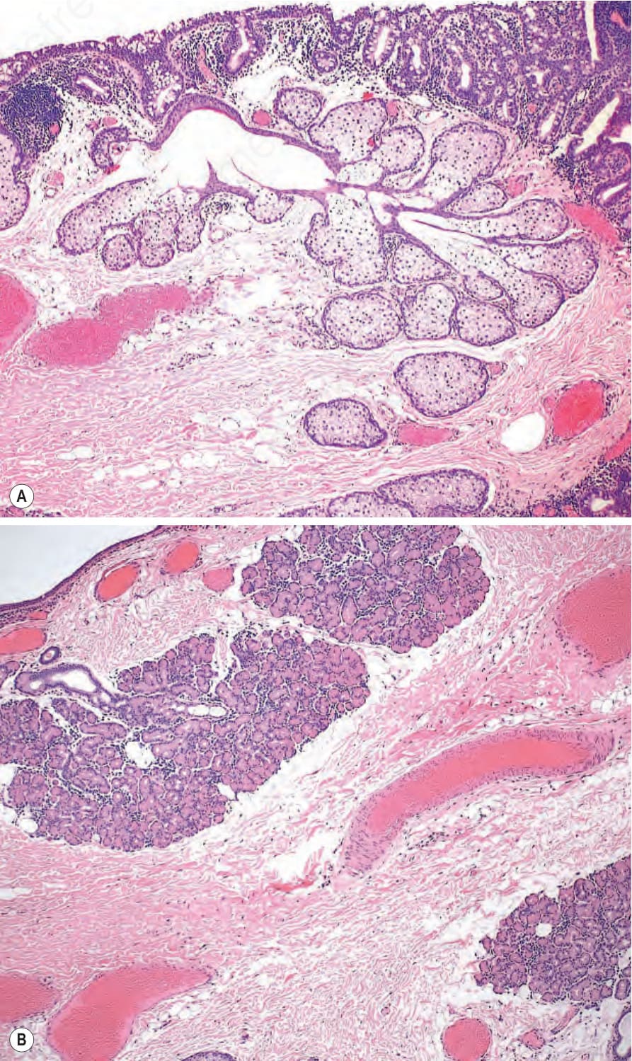
圖 27-9:複合型迷芽瘤 (complex choristoma):(A) 病灶的某一區域含有真皮型膠原 (dermal type collagen) 與脂肪組織 (adipose tissue),並有皮膚附屬器 (skin appendages)(皮脂腺 sebaceous glands)。(B) 在病灶的另一區域,可辨識出淚腺組織 (lacrimal gland tissue)。
Fig. 27.9 Complex choristoma: (A) one area of the lesion contained dermal type collagen and adipose tissue with skin appendages (sebaceous glands). (B) In a different area of the lesion, lacrimal gland tissue was identified.