Dermoid
Dermoid
Clinical features Epibulbar dermoid (Fig. 27.6) is a congenital lesion that is most commonly located inferotemporally at the limbus. It is well circumscribed, tan-yellow, and may have fine hair protruding from the surface. The lesion may be isolated, or associated with Goldenhar syndrome (oculoauriculovertebral dysplasia).1 Patients should be evaluated for preauricular skin appendages, hearing loss, eyelid colobomas, mandibular hypoplasia, and vertebral abnormalities.2 Dermoid can cause astigmatism and amblyopia.
Histologic features Complex choristomas may include a combination of ectopic tissues, including dermal tissue with adipose tissue and dermal appendages, lacrimal gland tissue, smooth muscle, cartilage, and bone (Figs 27.9 and 27.10).2
Histologic features Epibulbar dermoid is composed of dense fibrous tissue containing dermal appendages, such as hair follicles, sebaceous glands, sweat glands, and sometimes adipose tissue, lined by nonkeratinizing stratified squamous epithelium (Fig. 27.7).
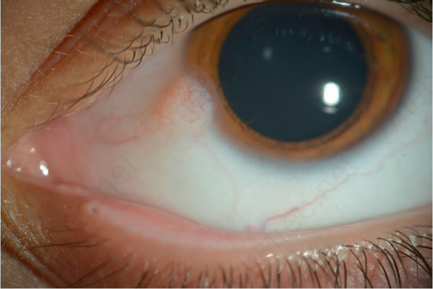
Fig. 27.6 Limbal dermoid: clinical photograph showing an elevated, yellow-white lesion at the limbus inferonasally.
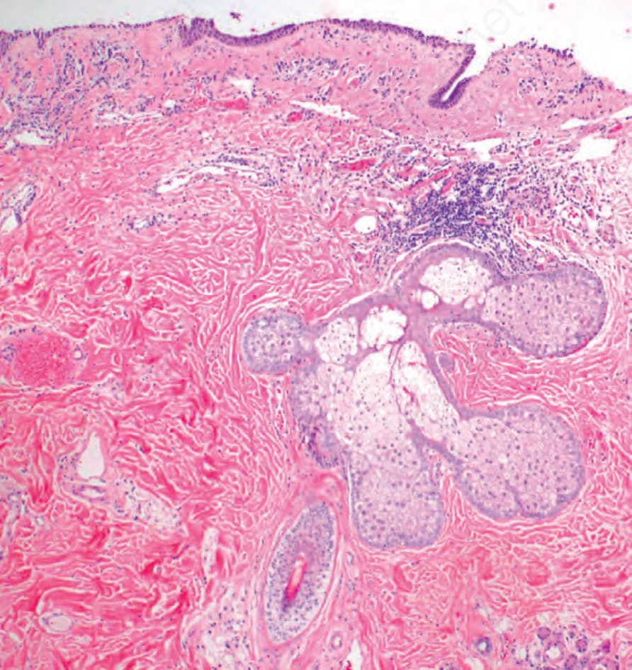
Fig. 27.7 Limbal dermoid: histology shows dermal type collagen and skin adnexal structures, including hair follicle and sebaceous gland.
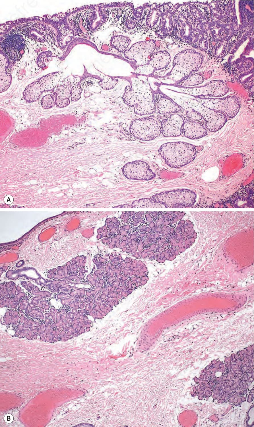
Fig. 27.9 Complex choristoma: (A) one area of the lesion contained dermal type collagen and adipose tissue with skin appendages (sebaceous glands). (B) In a different area of the lesion, lacrimal gland tissue was identified.