印戒細胞型黑色素瘤 (Signet Ring Cell Melanoma)
印戒細胞型黑色素瘤 (signet ring cell melanoma) 是一種極為罕見的組織學變異型,與 rhabdoid 變異型(見下文)有相當程度的重疊。它大多描述於轉移性腫瘤。亦有印戒細胞 (signet ring cell) 的相關報告。
「rhabdoid 腫瘤 (rhabdoid tumor)」一詞最初是用來描述一種侵襲性的兒童腎臟腫瘤,當時被認為代表 nephroblastoma 的一種橫紋肌肉瘤樣 (rhabdomyosarcomatous) 變異型。其特徵為存在嗜酸性玻璃樣胞質內包涵體 (eosinophilic hyaline intracytoplasmic inclusions),後來經超微結構鑑定為中間絲 (intermediate filaments) 的渦旋。腎外 rhabdoid 腫瘤 (extrarenal rhabdoid tumors) 已於眾多部位被發現,包括軟組織、皮膚、腦、眼眶、子宮、膀胱、攝護腺與肝臟。rhabdoid 表型 (rhabdoid phenotype) 最早是描述於轉移性黑色素瘤 (metastatic melanoma)。較近期,有少數呈現 rhabdoid 特徵的原發腫瘤被記載。目前文獻記載的病例過少,無法判定黑色素瘤中的 rhabdoid 表型是否具有任何預後意義。
rhabdoid 表型的特徵為存在大型上皮樣細胞 (epithelioid cells),具有豐富的胞質,內含一個大型圓形的嗜酸性玻璃樣包涵體 (eosinophilic hyaline inclusion)(Fig. 26.72)。細胞核典型為泡狀核 (vesicular),核仁則為嗜酸性且明顯。腎外 rhabdoid 腫瘤並非一個均質的實體;rhabdoid 特徵最常見的情形是局灶性地出現於一個原本可辨識且可分類的病灶中,亦即 rhabdoid 變化代表去分化 (dedifferentiation) 過程中的一種形態學終點。
免疫細胞化學 (immunocytochemistry) 的結果不一。部分腫瘤的特徵為表現 S100 protein、SOX10 與 HMB-45(Fig. 26.73)。Mart-1 亦常由腫瘤細胞表現。然而在若干腫瘤中,rhabdoid 細胞並不表現上述任何標記,且其包涵體含有 keratin、smooth muscle actin 或 desmin。罕見的 rhabdoid 形態黑色素瘤病例會呈現橫紋肌肉瘤樣分化 (rhabdomyosarcomatous differentiation),表現 desmin、myogenin 與 MYOD1。
較古典型黑色素瘤更常見。預後通常不佳,但這與就診時的腫瘤厚度 (tumor thickness) 有關,而非此一不尋常黑色素瘤變異型所特有的任何特徵。氣球細胞變化 (balloon cell change) 在 nevi 中似乎較黑色素瘤中更為常見。
在超微結構上,這些包涵體通常被發現由中間絲 (intermediate filaments) 的聚集所構成。在一組轉移性黑色素瘤的系列中,rhabdoid 包涵體是由擴張的粗糙內質網 (rough endoplasmic reticulum) 與粒線體 (mitochondria) 內的小管狀包涵體 (tubular inclusions) 所組成。
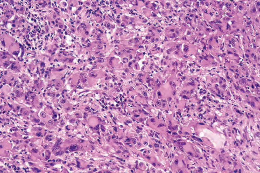
圖 26-72:Rhabdoid melanoma:此變異型常造成診斷困難,因為病灶通常為無黑色素 (amelanotic)。注意泡狀核 (vesicular nuclei)、明顯的核仁,以及大型嗜酸性胞質內包涵體。
Fig. 26.72 Rhabdoid melanoma: this variant often causes diagnostic difficulty since lesions are commonly amelanotic. Note the vesicular nuclei, prominent nucleoli, and large eosinophilic cytoplasmic inclusions.
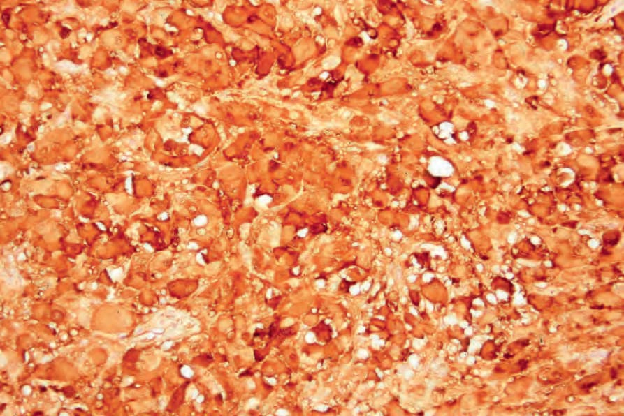
圖 26-73:Rhabdoid melanoma:在缺乏已知黑色素瘤病史的情況下,診斷通常仰賴免疫組織化學 (S100 protein)。
Fig. 26.73 Rhabdoid melanoma: in the absence of a known history of melanoma, the diagnosis commonly depends upon immunohistochemistry (S100 protein).
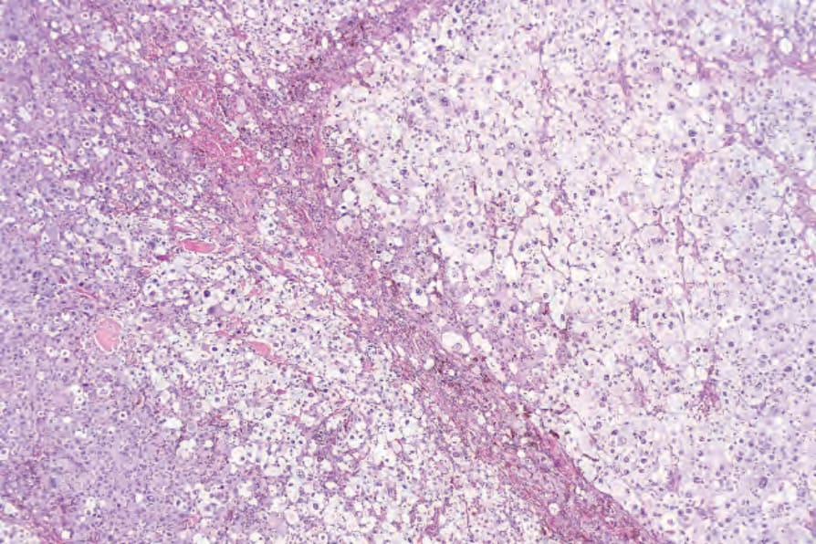
圖 26-74:Balloon cell melanoma:腫瘤細胞具有透明或輕微顆粒狀的胞質。此例為無黑色素 (amelanotic),很容易被誤認為黃色瘤樣 (xanthomatous) 病灶。
Fig. 26.74 Balloon cell melanoma: the tumor cells have clear or faintly granular cytoplasm. This example is amelanotic and could easily be mistaken for a xanthomatous lesion.
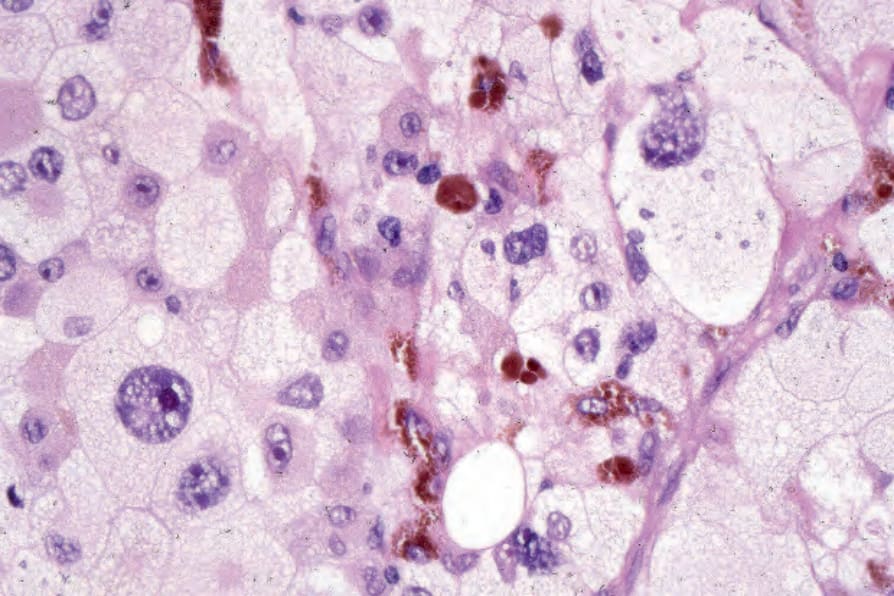
圖 26-75:Balloon cell melanoma:高倍視野。
Fig. 26.75 Balloon cell melanoma: high-power view.