Signet ring cell melanoma
Signet ring cell melanoma
Signet ring cell melanoma is a very rare histologic variant, which shows considerable overlap with rhabdoid variants (see below). It has been mostly described in metastatic tumors. There are also reports of signet ring cell
The term ‘rhabdoid tumor’ was originally coined to describe an aggressive childhood renal tumor, which was thought to represent a rhabdomyosarcomatous variant of nephroblastoma.2 It was characterized by the presence of eosinophilic hyaline intracytoplasmic inclusions, later identified ultrastructurally as whorls of intermediate filaments. Extrarenal rhabdoid tumors have been identified at a wide range of sites including soft tissues, skin, brain, orbit, uterus, bladder, prostate, and liver. A rhabdoid phenotype was first described in metastatic melanoma.3–10 More recently, small numbers of primary tumors showing rhabdoid features have been documented.11–17 At present, there are too few cases documented to determine whether the rhabdoid phenotype in melanoma has any prognostic significance.
The rhabdoid phenotype is characterized by the presence of large epithelioid cells with abundant cytoplasm containing a large round eosinophilic hyaline inclusion (Fig. 26.72). The nuclei are typically vesicular and nucleoli are eosinophilic and conspicuous. Extrarenal rhabdoid tumors are not a
1337 Histologic variants of melanoma
homogeneous entity; most often rhabdoid features are seen focally in an otherwise recognizable and classifiable lesion, i.e., rhabdoid change represents a morphological endpoint in dedifferentiation.
The results of immunocytochemistry are variable. Some tumors are characterized by S100 protein, SOX10, and HMB-45 expression (Fig. 26.73).11,12 Mart-1 is also often expressed by tumor cells. In a number of tumors, however, the rhabdoid cells fail to show any of the above and the inclusions contain keratin, smooth muscle actin, or desmin.3,12 Rare examples of melanoma with rhabdoid morphology display rhabdomyosarcomatous differentiation with expression of desmin, myogenin, and MYOD1.
is more commonly seen than in classic melanoma.2 Prognosis is usually poor but this relates to the tumor thickness at presentation rather than any feature specific to this unusual melanoma variant. Balloon cell change appears to be more common in nevi than melanoma.6
Ultrastructurally, the inclusions have usually been found to consist of aggregates of intermediate filaments. In one series of metastatic melanoma, the rhabdoid inclusions were composed of tubular inclusions within dilated rough endoplasmic reticulum and mitochondria.5
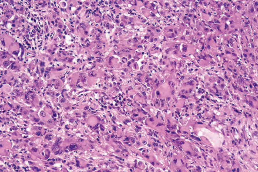
Fig. 26.72 Rhabdoid melanoma: this variant often causes diagnostic difficulty since lesions are commonly amelanotic. Note the vesicular nuclei, prominent nucleoli, and large eosinophilic cytoplasmic inclusions.
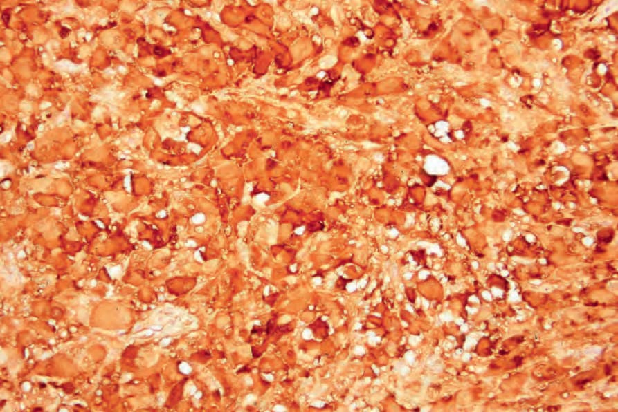
Fig. 26.73 Rhabdoid melanoma: in the absence of a known history of melanoma, the diagnosis commonly depends upon immunohistochemistry (S100 protein).
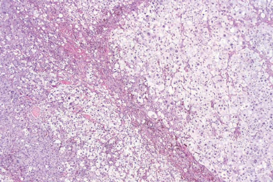
Fig. 26.74 Balloon cell melanoma: the tumor cells have clear or faintly granular cytoplasm. This example is amelanotic and could easily be mistaken for a xanthomatous lesion.
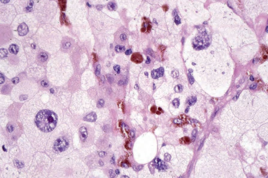
Fig. 26.75 Balloon cell melanoma: high-power view.