Hori nevus
Hori nevus
臨床特徵 (Clinical Features)
- Hori 痣 (Hori nevus)(亦稱 nevus fuscoceruleus zygomaticus、顏面與四肢之後天性真皮黑色素細胞增多症 (acquired dermal melanocytosis of the face and extremities)、後天性雙側 Ota 痣樣斑 (acquired bilateral nevus of Ota-like macules))是一種罕見的後天性雙側真皮黑色素細胞增多症 (dermal melanocytosis),主要侵犯亞洲女性。患者多在三、四十歲(第三與第四個十年)。早期病灶以散在的棕色斑 (brown macules) 為特徵,隨時間逐漸變得較為融合並轉為板岩灰色 (slate gray)。病灶一般不會自發性消退。
- Hori nevus 好發於臉頰的顴部 (malar region),其次為前額、上眼瞼、顳部,以及鼻根/鼻翼 (root/alae of the nose)。病灶也可發生於顏面以外的部位。在男性,最常見的發生部位是前額,且額外出現顏面外病灶 (extrafacial lesions) 的發生率較高。雖然最常由日曬與懷孕所誘發,其他因素還包括荷爾蒙藥物 (hormonal medications)、壓力與外傷。
- 近期一項前瞻性研究顯示,42% 的患者在一等親 (first-degree relatives) 中有陽性家族史。Hori nevus 亦曾被報告發生於頑固性濕疹 (refractory eczema) 及經治療之乾癬 (treated psoriasis) 的部位。曾有單一病例報告出現黏膜侵犯 (mucosal involvement)。
組織病理特徵 (Histopathology)
- 其特徵為一種表淺真皮樹突狀黑色素細胞增多症 (superficial dermal dendritic melanocytosis)。雙極樹突狀黑色素細胞 (bipolar dendritic melanocytes) 散布於真皮上層,常與表皮平行排列,並呈血管周圍分布 (perivascular distribution)。
1295 真皮黑色素細胞病灶(真皮黑色素細胞增多症)(Dermal melanocytic lesions, dermal melanocytoses)
組織病理特徵 (Histologic features) 普通藍痣 (common blue nevus) 典型上位於網狀真皮 (reticular dermis) 的較深部,但偶爾可出現於表淺真皮,或從乳頭真皮 (papillary dermis) 延伸至皮下脂肪 (Fig. 25.227)。雖然其上方的表皮通常正常,但有時也會合併接合處活性 (junctional activity)(或一個良性的真皮內成分),即合併痣 (combined nevus)(見上文)。其組成為一群常為高度色素沉著的雙極樹突狀梭形細胞 (bipolar, dendritic spindled cells),伴隨宿主來源的緻密纖維母細胞與膠原性梭形細胞反應 (fibroblastic and collagenous spindled cell response),並常伴有高度色素沉著的噬黑色素細胞 (melanophages)(Figs 25.228 與 25.229)。有絲分裂相 (mitotic figures) 罕見,且無多形性 (pleomorphism)。這些黑色素細胞常在皮膚附屬器、血管與神經周圍形成聚集,並常與表面上皮平行排列。整體而言,common blue nevus 的浸潤密度遠較 Ito 痣或 Ota 痣 (nevi of Ito or Ota) 為高。曾有罕見病例報告於 common/combined blue nevus 內出現平滑肌增生 (smooth muscle hyperplasia)。
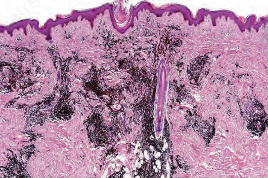
圖 25-227:普通藍痣 (common blue nevus):此高度色素沉著的梭形細胞腫瘤 (spindled cell neoplasm) 廣泛侵犯網狀真皮 (reticular dermis)。
Fig. 25.227 Common blue nevus: this highly pigmented spindled cell neoplasm extensively involves the reticular dermis.
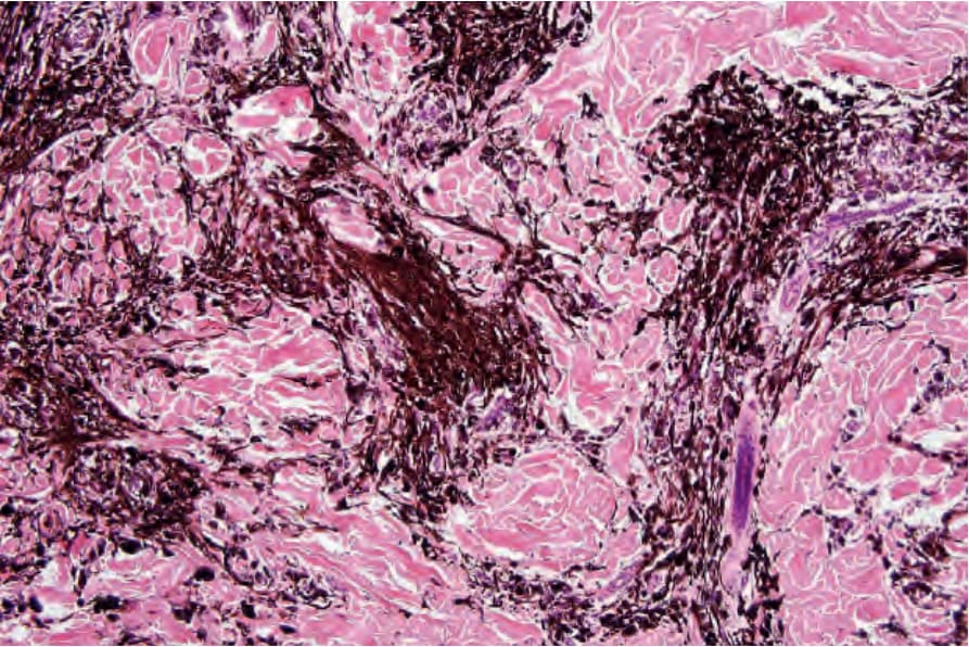
圖 25-228:普通藍痣 (common blue nevus):中倍視野 (medium-power view)。
Fig. 25.228 Common blue nevus: medium-power view.