毛髮週期 (Hair Cycle)
毛髮的生長週期,無論是終毛 (terminal) 或毳毛 (vellus),皆分為三個階段:活躍生長期 (anagen)、退化期 (catagen)、休止期 (telogen);而作為休止期最後階段的延伸,另有兩個額外階段:毛幹脫出期 (hair shaft-extrusion,exogen) 與空毛囊期 (empty hair follicle,kenogen)(圖 22.25)。anagen 期的持續時間決定了毛幹的長度。人類與其他哺乳動物不同,這些階段是連續性發生而非同步性發生。覆蓋於體表的毛髮並無週期性的更替。每個階段的持續時間與相對比例各不相同:anagen 期 2–7 年(百分之八十五至百分之一百)、catagen 期 2–3 週(百分之零至百分之一)、telogen 期 100 天(百分之零至百分之十五)。微小化毛囊 (miniaturized hair follicles) 與毳毛的週期快得多,因而縮短了 anagen 期。
眾多生長因子、細胞激素 (cytokines)、荷爾蒙、神經傳導物質及其受體之間的交互作用,對正常毛囊的發育與週期循環十分重要。週期循環的驅動力,即「毛髮週期時鐘 (hair cycle clock)」,似乎位於毛囊本身、其鄰近的微環境 (niche) 以及真皮的微環境中。然而,並無任何單一生長因子對這些過程具有最終的控制作用。
anagen 期持續 2 至 7 年,其特徵為持續生長,產生一根長而帶有色素、容易看見的毛幹 (pilar stem)。它是決定毛髮長度的階段,也是依身體部位不同而變化最大的階段。在形態上,它對應於前述深位於皮下脂肪中的終毛毛囊(見圖 22.19)。處於 anagen 期的毛囊其毛球 (pilar bulb) 顯示出大量黑色素生成與旺盛的有絲分裂活性,使毛髮約以每月 1.0 公分的速度生長。在任一特定時刻,有百分之八十五至百分之一百的毛囊處於 anagen 期。由於此期具有最高的有絲分裂活性、黑色素生成 (melanogenesis) 及 DNA 合成,因此最易受荷爾蒙變化、藥物及各種毒素的影響。
毛髮的色素生成僅發生於 anagen 期,由毛球部黑色素細胞 (bulbar melanocytes)、角質形成細胞 (keratinocytes) 與真皮乳頭纖維母細胞 (dermal papilla fibroblasts) 之間的交互作用所誘發(此即 hair follicle pigmentary unit,毛囊色素單位)(見圖 22.22)。毛囊黑色素生成與 anagen 期的耦合,使毛囊黑色素生成有別於表皮的持續性黑色素生成。hair follicle pigmentary unit 的週期性重建僅在最初 10 個毛髮週期內達到最佳狀態,亦即直到約 40 歲為止。目前尚不清楚毛髮變白是功能喪失的結果,抑或是人類毛髮黑色素細胞選擇性耗竭的結果。
catagen 期位於 telogen 期之前,在此階段中,毛囊的下段 (inferior segment) 經歷大量凋亡 (apoptosis),使其體積縮小。它是最短的階段,持續 2 至 3 週。僅有百分之一至百分之二的毛囊處於 catagen 期,且在正常頭皮切片中很少見到。水平切面的特徵為瀰漫性凋亡,以及缺乏有絲分裂與色素活性。凋亡細胞具有強烈嗜伊紅性的細胞質,並有深染的核固縮 (pyknotic) 細胞核(圖 22.26 與圖 22.27)。隨著毛囊退縮,玻璃膜 (vitreous membrane) 塌陷,並呈現增厚與皺褶狀的外觀(圖 22.28)。重要的是不要將此現象與盤狀紅斑性狼瘡 (discoid lupus) 中所見的基底膜 (basal membrane) 增厚相混淆。內毛根鞘 (inner root sheath) 消失,外毛根鞘 (outer root sheath) 在形態上類似於峽部 (isthmus) 的上皮,圍繞著一根杵狀 (club-shaped) 的毛幹,並以毛鞘 (trichilemmal) 方式角化(圖 22.29)。catagen 期毛囊常見於拔毛癖 (trichotillomania)。
由於 catagen 期短暫且很快接續 telogen 期,因此就量化目的而言,catagen 期與 telogen 期的毛囊會合併計算。telogen 期持續 100 天。在任一特定時刻,約有百分之十至百分之十五的毛髮總數處於 telogen 期,每天約有 100 根 telogen 期毛髮脫落。處於 telogen 期的毛囊代表下段退化的最終階段。毛乳頭 (hair papilla) 位於豎毛肌 (arrector pili muscle) 附著區的下方,在水平切面中呈現為一團基底樣細胞 (basaloid cells)(即次級胚芽 (secondary germ):telogen germinal unit,休止期生發單位)(圖 22.30 與圖 22.31)。過去認為毛囊幹細胞 (hair follicle stem cells) 位於 telogen germinal unit。然而,真正的幹細胞位於膨大部 (bulge),即豎毛肌的附著點;這些細胞不僅負責再生毛囊,也負責再生皮脂腺與表皮(圖 22.32)。在人類毛囊中,膨大部很少可見,因此其偵測需借助免疫組織化學染色 (immunohistochemistry,cytokeratin 15)。膨大部幹細胞的喪失會導致瘢痕性禿髮 (cicatricial types of alopecia)。
毛囊也包含皮脂腺 (sebaceous glands),其分泌物潤滑毛道與皮膚表面。它們數量眾多,在上段 (superior segment) 層級的切片中很容易辨識。瘢痕性禿髮 (scarring alopecias) 的一個早期現象即為皮脂腺的喪失。在特定部位,如腋窩、生殖器區、腹部中央與乳暈,頂泌汗腺 (apocrine glands) 也與毛囊相關聯。
毛囊下段完全退化,僅留下一個稱為毛囊星狀體 (follicular stella;又稱 follicular streamer、fibrous streamer,纖維束帶) 的結構,標示出已退縮毛囊先前所在的位置(圖 22.33)。follicular stella 並非 telogen 期所特有,因為在雄性禿 (androgenetic alopecia) 中終毛毛囊微小化所造成的結果中也可見到它。與瘢痕性禿髮中所見者不同,這些 follicular stella 在偏光下不具雙折射性 (birefringent)(圖 22.34)。
另有兩個階段被視為 telogen 階段的最終組成部分:
- exogen(teloptosis,毛髮脫落)代表一根 telogen 期杵狀毛 (telogen club),它已失去杵狀毛髮細胞與其上皮包膜細胞之間的黏附,顯然是經由某種蛋白水解機制活化了一個主動的脫落過程。此即每日脫落的 100 根毛髮。
- kenogen 指的是 telogen 期毛囊脫落後留下的一個空毛囊。處於 exogen 與 kenogen 的毛囊在組織學上不易分類。
毛囊免疫豁免 (Hair follicle immune privilege) 毛囊免疫系統具有獨特的構造,使其在毛囊週期的 anagen 階段維持一個相對免疫豁免 (relative immune privilege) 的區域。毛囊近端上皮(內毛根鞘與毛基質 hair matrix)的特徵為:主要組織相容性複合體第一型 a 類抗原 (major histocompatibility complex class Ia antigens) 的表現量極低、局部產生免疫抑制因子(transforming growth factor [TGF]-beta 1、alpha-melanocyte-stimulating hormone)、以及抑制自然殺手細胞 (natural killer,NK) 的活性。此免疫豁免的崩潰,可能在圓禿 (alopecia areata) 的致病機轉中扮演重要角色,並可能也涉及某些瘢痕性禿髮。
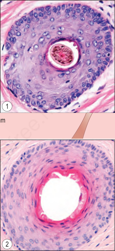
圖 22-16:終毛 anagen 期,上段的垂直與水平切面:注意毛囊口 (ostium)、漏斗部 (infundibulum) 與峽部 (isthmus)。插圖顯示通過 (1) infundibulum 與 (2) isthmus 的水平切面。infundibulum 的外毛根鞘 (outer root sheath) 由類似表皮的鱗狀上皮組成。Courtesy of M. Mejia, MD, Universidad Pontificia Bolivariana, Medellín, Colombia.
Fig. 22.16 Terminal anagen hair, vertical and horizontal sections of the upper segment: note the ostium, infundibulum and isthmus. The insets show horizontal sections through (1) the infundibulum and (2) the isthmus. The outer root sheath of the infundibulum is composed of squamous epithelium similar to the epidermis. Courtesy of M. Mejia, MD, Universidad Pontificia Bolivariana, Medellín, Colombia.
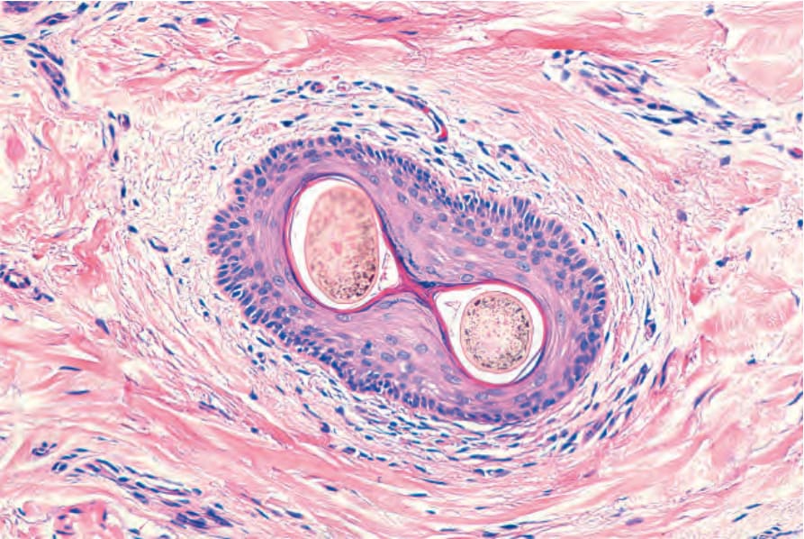
圖 22-17:漏斗部 (infundibulum)。水平切面。其壁由鱗狀複層角化上皮 (squamous stratified keratinized epithelium) 構成。周圍真皮被一些淋巴球浸潤。有兩根毛幹從單一毛囊口 (ostium) 中冒出。
Fig. 22.17 Infundibulum. Horizontal section. The wall is made up of squamous stratified keratinized epithelium. The surrounding dermis is infiltrated by some lymphocytes. There are two hair shafts emerging from a single ostium.
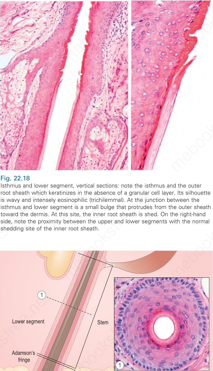
圖 22-18:峽部 (isthmus) 與下段,垂直切面:注意 isthmus 與在缺乏顆粒細胞層 (granular cell layer) 情況下角化的外毛根鞘。其輪廓呈波浪狀且強烈嗜伊紅性 (trichilemmal,毛鞘性)。在 isthmus 與下段交界處有一個從外鞘向真皮突出的小膨大部 (bulge)。在此處,內毛根鞘脫落。在右側,注意上段與下段之間的鄰近關係,以及內毛根鞘的正常脫落部位。
Fig. 22.18 Isthmus and lower segment, vertical sections: note the isthmus and the outer root sheath which keratinizes in the absence of a granular cell layer. Its silhouette is wavy and intensely eosinophilic (trichilemmal). At the junction between the isthmus and lower segment is a small bulge that protrudes from the outer sheath toward the dermis. At this site, the inner root sheath is shed. On the right-hand side, note the proximity between the upper and lower segments with the normal shedding site of the inner root sheath.
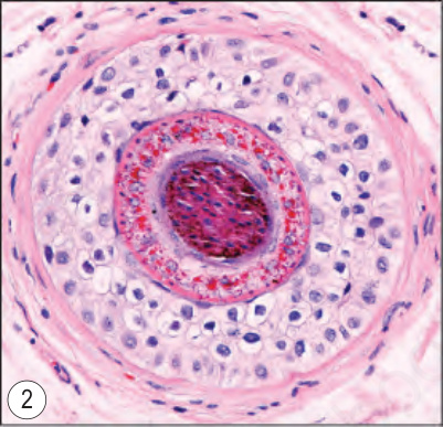
圖 22-19:終毛 anagen 期,下段的垂直與水平切面:注意毛幹與位於 Adamson fringe (Adamson 緣) 深部的毛球 (hair bulb)。水平切面顯示 (1) 毛幹 (stem) 與 (2) 毛球上部。注意毛幹與內毛根鞘中細胞核的消失。Courtesy of M. Mejia, MD, Universidad Pontificia Bolivariana, Medellín, Colombia.
Fig. 22.19 Terminal anagen hair, vertical and horizontal sections of the lower segment: note the shaft and the hair bulb deep to Adamson fringe. The horizontal sections show (1) the stem and (2) the upper part of the bulb. Note the loss of nuclei in the hair shaft and the inner root sheath. Courtesy of M. Mejia, MD, Universidad Pontificia Bolivariana, Medellín, Colombia.
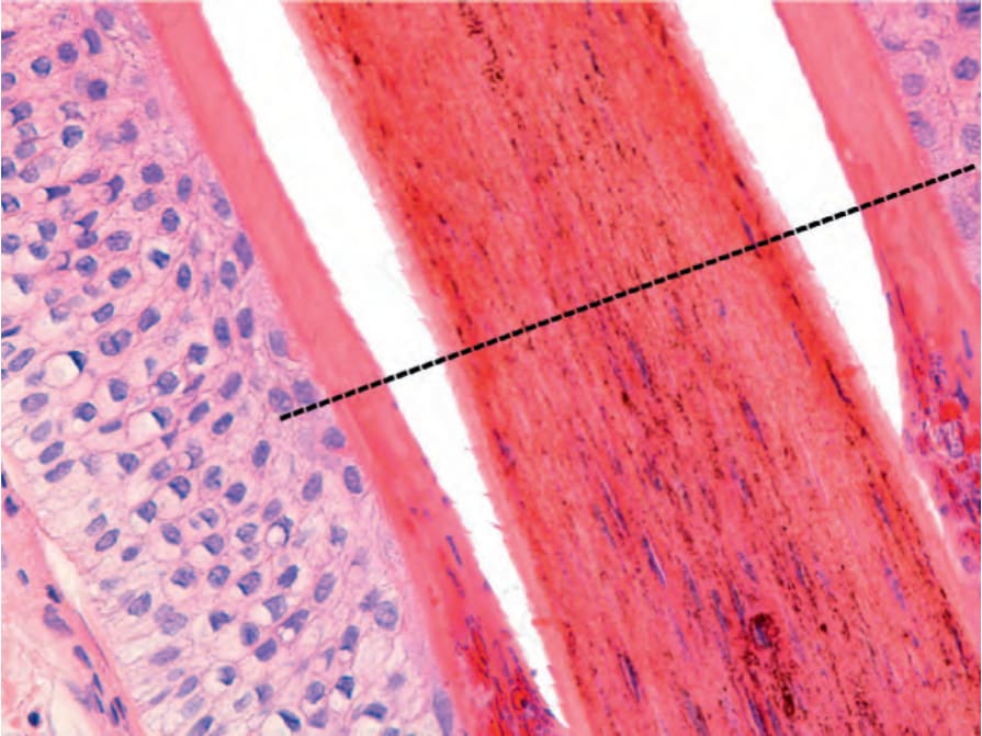
圖 22-20:顯示 Adamson fringe (Adamson 緣) 的終毛 anagen 期切面:在毛幹 (stem,上方) 與毛球 (bulb,下方) 交界處,內毛根鞘與毛幹中喪失了毛透明蛋白顆粒 (trichohyalin granules) 與細胞核。在此層級以上,內毛根鞘完全角化。
Fig. 22.20 Terminal anagen hair section showing Adamson fringe: there is loss of trichohyalin granules and nuclei in the inner root sheath and the hair shaft at the junction of the stem (above) and the bulb (below). Above this level, the inner root sheath keratinizes completely.
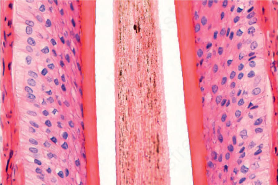
圖 22-21:終毛 anagen 期,下段:在此垂直切面中,粉紅色的內毛根鞘 (無核) 與外毛根鞘 (有核) 形成明顯對比。角皮 (cuticle) 具有鋸齒狀邊緣,其方向與內毛根鞘的相反。外毛根鞘的細胞因在此層級具有顯著的細胞質內肝醣 (intracytoplasmic glycogen) 而具有透明的細胞質。
Fig. 22.21 Terminal anagen hair, lower segment: the pink inner root sheath (anucleated) clearly contrasts with the outer root sheath (nucleated) in this vertical section. The cuticle has a serrated border orientated in the opposite direction to that of the inner root sheath. The cells of the outer root sheath have clear cytoplasm due to prominent intracytoplasmic glycogen at this level.
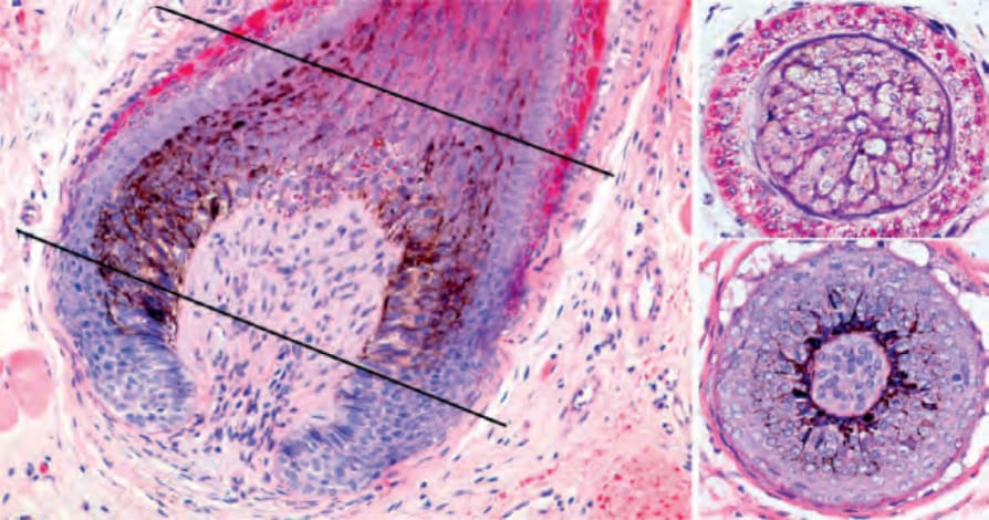
圖 22-22:終毛 anagen 期:毛球與 hair follicle pigmentary unit (毛囊色素單位),垂直與水平切面。在垂直與水平切面中,注意基質上層細胞 (supramatricial cells) 具有由圍繞真皮乳頭的樹突狀黑色素細胞 (dendritic melanocytes) 所轉移來的細胞質內色素。後者 (真皮乳頭) 由結締組織與血管構成。
Fig. 22.22 Terminal anagen hair: hair bulb and hair follicle pigmentary unit, vertical and horizontal sections. In the vertical and horizontal sections, note the supramatricial cells with intracytoplasmic pigmentation transferred from the dendritic melanocytes that surround the dermal papilla. The latter is composed of connective tissue and blood vessels.
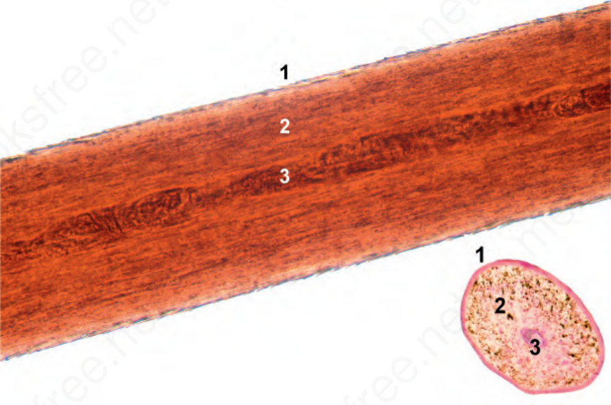
圖 22-23:毛幹 (hair shaft):注意皮質 (cortex,2) 與角皮 (cuticle,1)。有時,髓質 (medulla,3) 很容易看見。
Fig. 22.23 Hair shaft: Note the cortex (2) and the cuticle (1). Sometimes, the medulla is easily visualized (3).
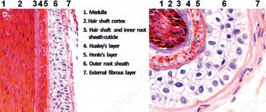
圖 22-24:終毛 anagen 期毛囊:緊鄰 Adamson fringe (Adamson 緣) 下方的水平與垂直切面。注意毛囊的不同層次。(1) 髓質 (medulla)、(2) 皮質 (cortex)、(3) 毛髮角皮與內毛根鞘角皮 (cuticle of the hair and cuticle of the inner root sheath)、(4) Huxley layer (Huxley 層)、(5) Henle layer (Henle 層)、(6) 外毛根鞘 (outer root sheath)、以及 (7) 玻璃膜與外纖維層 (vitreous and external fibrous layer,毛囊周圍結締組織鞘 perifollicular connective tissue sheath)。
Fig. 22.24 Terminal anagen hair follicles: horizontal and vertical sections immediately below the Adamson fringe. Note the different layers of the hair follicle. (1) Medulla, (2) cortex, (3) cuticle of the hair and cuticle of the inner root sheath, (4) Huxley layer, (5) Henle layer, (6) outer root sheath, and (7) vitreous and external fibrous layer (perifollicular connective tissue sheath).
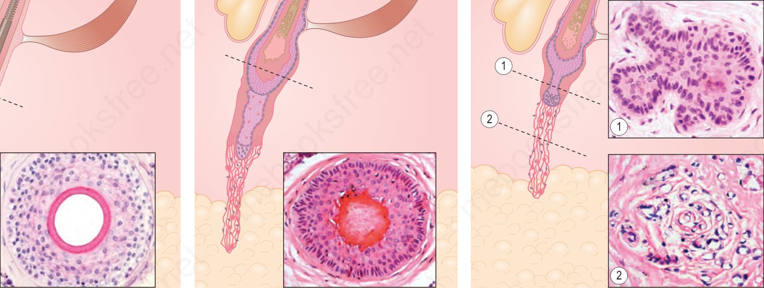
圖 22-25:(A、B) 毛囊週期 (hair follicle cycle):毛囊由 anagen 持續循環至 catagen、telogen 與 exogen,再經由下段的生長返回 anagen。每個階段的持續時間與相對比例各不相同。
Fig. 22.25 (A, B) Hair follicle cycle: the hair follicle cycles continuously from anagen to catagen, telogen, and exogen, returning to anagen through growth of the lower segment. The duration and relative proportion of each stage is variable.
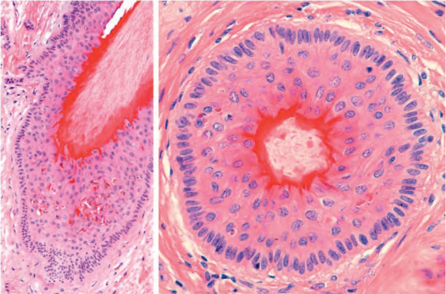
圖 22-26:catagen 期的終毛毛囊,垂直與水平切面:注意毛囊的退化以及圍繞杵狀毛幹的深紅色 trichilemmal keratinization (毛鞘性角化)。外毛根鞘顯示明顯的凋亡。在毛囊最深部,可見次級胚芽 (secondary germ) 形成的徵兆。
Fig. 22.26 Terminal hair follicle in catagen, vertical and horizontal sections: note the involution of the follicle and the deep red trichilemmal keratinization around the club-shaped hair shaft. The outer root sheath shows marked apoptosis. At the deepest part of the follicle, there is a hint of secondary germ formation.
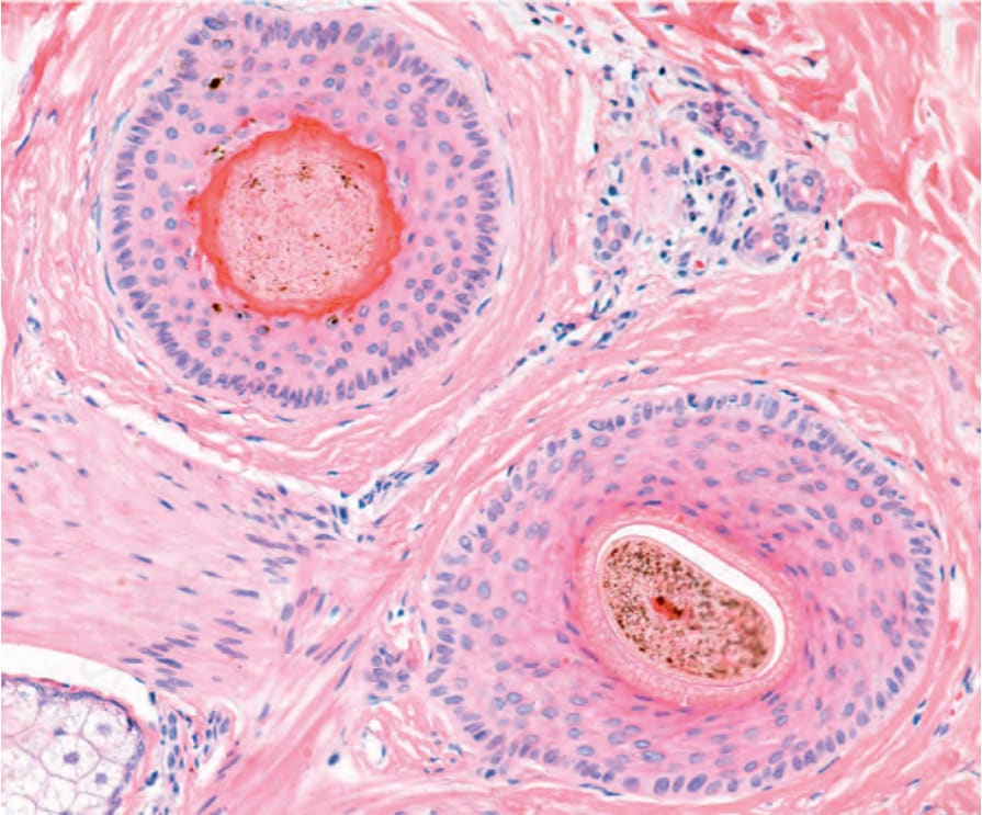
圖 22-27:catagen 期與 anagen 期的終毛毛囊,水平切面:注意 catagen 期毛囊 (左上) 與 anagen 期重度色素沉著終毛毛囊 (下方) 之間的差異——前者具有圍繞毛幹的 trichilemmal keratin (毛鞘性角質)、凋亡細胞與黑色素喪失。
Fig. 22.27 Terminal hair follicles in catagen and anagen, horizontal sections: note the differences between a catagen follicle (top left) with trichilemmal keratin surrounding the hair shaft, apoptotic cells and loss of melanin and an anagen, heavily pigmented terminal follicle (inferior).
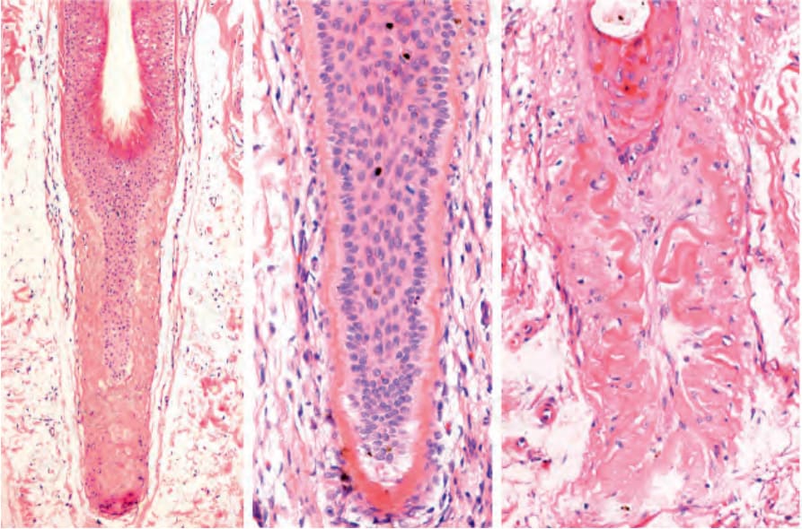
圖 22-28:catagen 期與 telogen 期的終毛毛囊,垂直切面:毛囊的退化遺留下一個增厚的基底膜 (basement membrane)。
Fig. 22.28 Terminal hair follicles in catagen and telogen, vertical sections: the involution of the hair follicle leaves behind a thickened basement membrane.
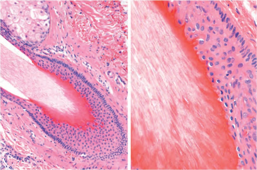
圖 22-29:晚期 catagen 期的終毛毛囊:外毛根鞘已喪失,且角化為 trichilemmal (毛鞘性) 類型。
Fig. 22.29 Terminal hair follicles in late catagen: the outer root sheath has been lost and the keratinization is of trichilemmal type.
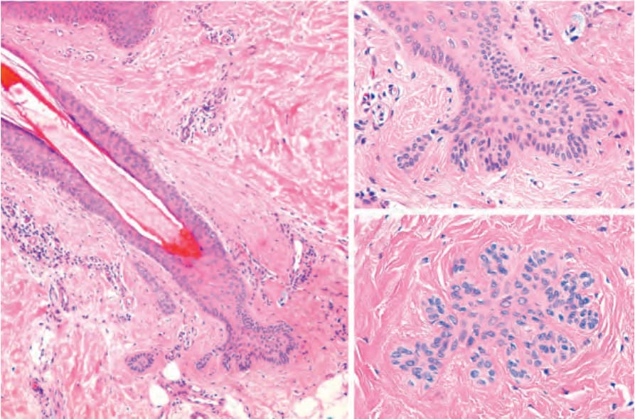
圖 22-30:telogen 期的終毛毛囊,次級胚芽 (secondary germ):在水平切面中,注意基底樣細胞凝聚成星狀 (asterisk) 或雛菊狀 (daisy shape) 的構造。
Fig. 22.30 Terminal hair follicle in telogen, secondary germ: note the condensation of basaloid cells in an asterisk or daisy shape in a horizontal section.
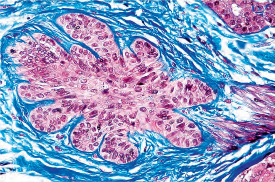
圖 22-31:telogen 期的終毛毛囊,次級胚芽 (secondary germ):以 Masson trichrome (Masson 三色染色) 染色的水平切面。
Fig. 22.31 Terminal hair follicle in telogen, secondary germ: horizontal section stained with Masson trichrome.
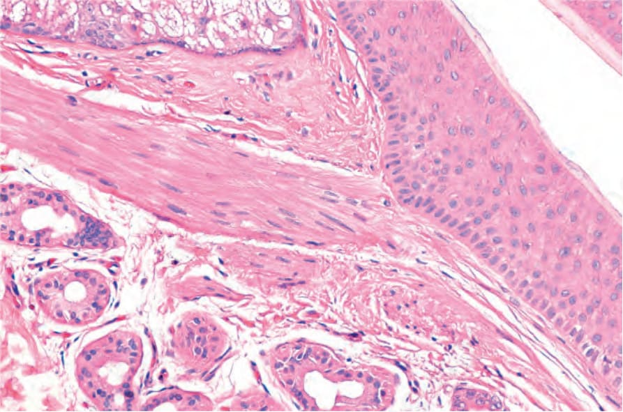
圖 22-32:膨大部 (bulge) 與豎毛肌 (arrector pili muscle)。在此垂直切面中,注意豎毛肌附著於膨大部的情形。
Fig. 22.32 Bulge and arrector pili muscle. In this vertical section, note the insertion of the arrector pili muscle erector in the bulge.
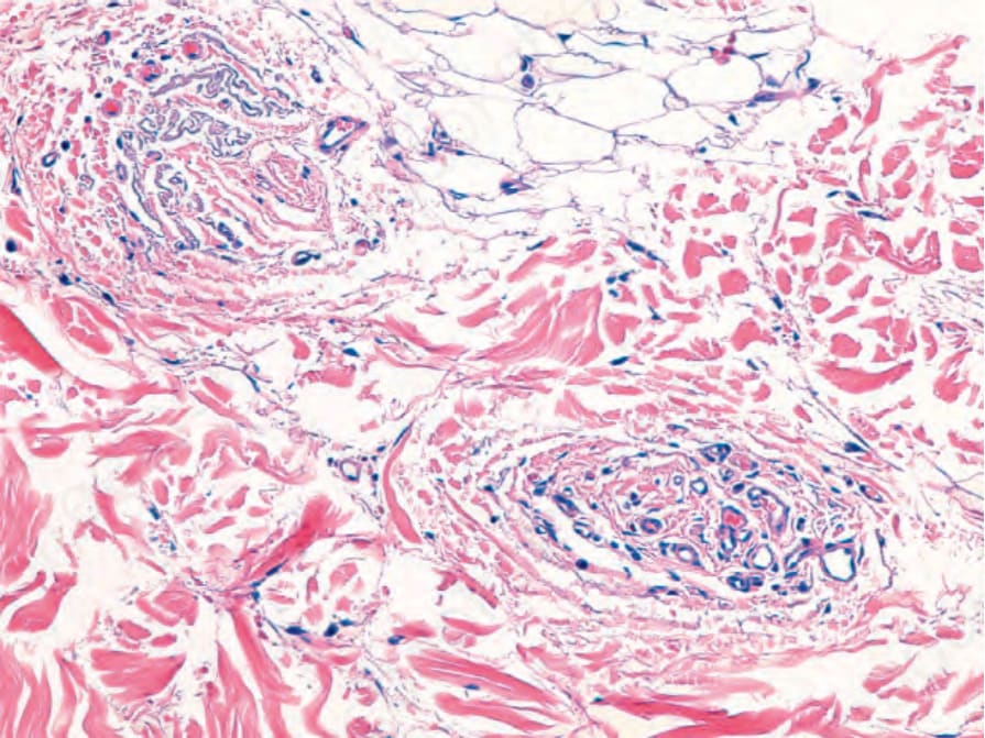
圖 22-33:毛囊星狀體 (follicular stella),水平切面:注意小血管與同心圓排列的結締組織相混合。
Fig. 22.33 Follicular stella, horizontal section: note the small blood vessels mixed with concentrically arranged connective tissue.
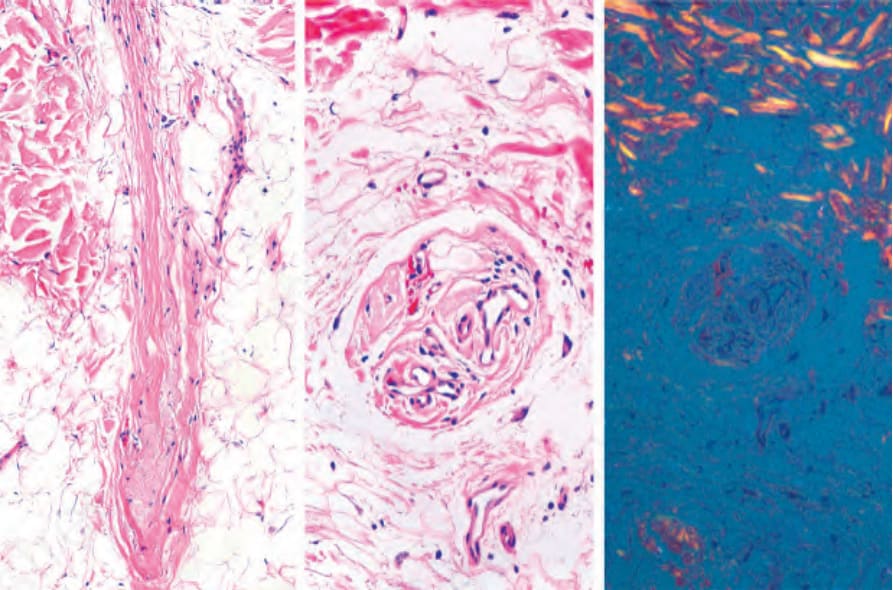
圖 22-34:毛囊星狀體 (follicular stella):在垂直與水平切面中,注意由膠原纖維 (collagen fibers)、纖維母細胞 (fibroblasts) 與血管組成的纖維血管性星狀體。在膠原纖維之間可見彈力組織變性物質 (elastotic material)。在偏光下,follicular stellae 不具雙折射性 (birefringence)。
Fig. 22.34 Follicular stella: note the fibrovascular stella composed of collagen fibers, fibroblasts, and blood vessels in vertical and horizontal sections. The elastotic material is apparent between the collagen fibers. With polarized light, there is no birefringence of the follicular stellae.