幼年性黃色肉芽腫 (Juvenile Xanthogranuloma)
幼年性黃色肉芽腫 (juvenile xanthogranuloma)
此病灶通常侵犯頭部與軀幹,但也可能侵犯生殖器 (Fig. 12.281)。1 已有文獻記載多灶性陰莖表現,2 以及單發性會陰丘疹3 與陰囊腫脹。4 臨床病理上完全相同的單發性病灶亦可見於成人。5 組織學上為富含脂質的組織球 (lipid-laden histiocytes) 與巨細胞 (giant cells),對 CD1 與 langerin 為陰性——此二者見於 Langerhans 細胞組織球增生症 (Langerhans cell histiocytosis)。約 25% 的病例 S100 可能為陽性。Juvenile xanthogranuloma 與異常脂質無關,但可能與色素性蕁麻疹 (urticaria pigmentosa)、糖尿病 (diabetes mellitus)、神經纖維瘤病 (neurofibromatosis)、巨細胞病毒感染 (cytomegalovirus infection) 及白血病 (leukemia) 有關聯。
另有一例孤立性陰蒂病灶的報告。6
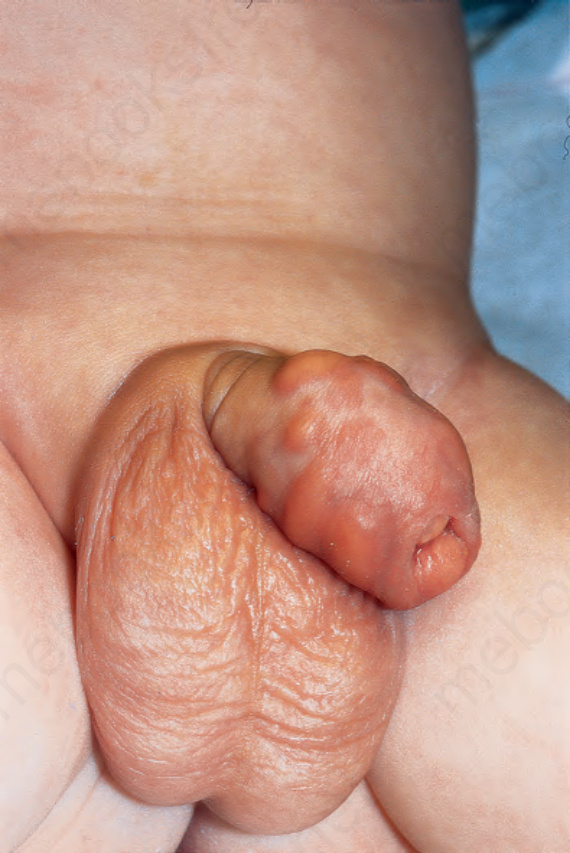
圖 12-281:幼年性黃色肉芽腫 (juvenile xanthogranuloma):病灶位於陰莖外側幹部。Courtesy of R. Haufmann, Ulm, Germany. Reproduced with permission from Haufmann R.E., Bachor, R. Juvenile xanthogranuloma of the penis. J Urol. 1993:150:456–457. From Bunker C. Male Genital Skin Disease. Saunders Ltd./Elsevier 2004.
Fig. 12.281 Juvenile xanthogranuloma: lesions are present on the lateral shaft of the penis. Courtesy of R. Haufmann, Ulm, Germany. Reproduced with permission from Haufmann R.E., Bachor, R. Juvenile xanthogranuloma of the penis. J Urol. 1993:150:456–457. From Bunker C. Male Genital Skin Disease. Saunders Ltd./Elsevier 2004.
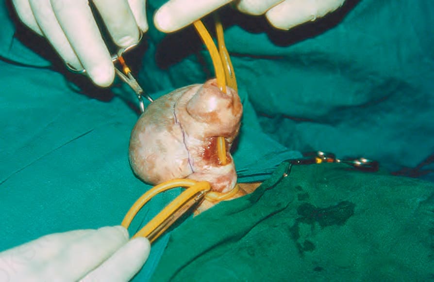
圖 12-282:慢性水腫類似蟹足腫 (chronic edema simulating a keloid):注意背側近端腫脹與腹側尿道瘻管 (ventral urethral fistula)。Courtesy of Dr Rameshwar Bang, Safat, Kuwait. Reproduced from Bang R.L. Penile edema induced by continuous condom catheter use and mimicking keloid scar. Scand J Urol Nephrol 1994;28:333–5. From Bunker C. Male Genital Skin Disease. Saunders Ltd./Elsevier 2004.
Fig. 12.282 Chronic edema simulating a keloid: note the dorsal proximal swelling and ventral urethral fistula. Courtesy of Dr Rameshwar Bang, Safat, Kuwait. Reproduced from Bang R.L. Penile edema induced by continuous condom catheter use and mimicking keloid scar. Scand J Urol Nephrol 1994;28:333–5. From Bunker C. Male Genital Skin Disease. Saunders Ltd./Elsevier 2004.
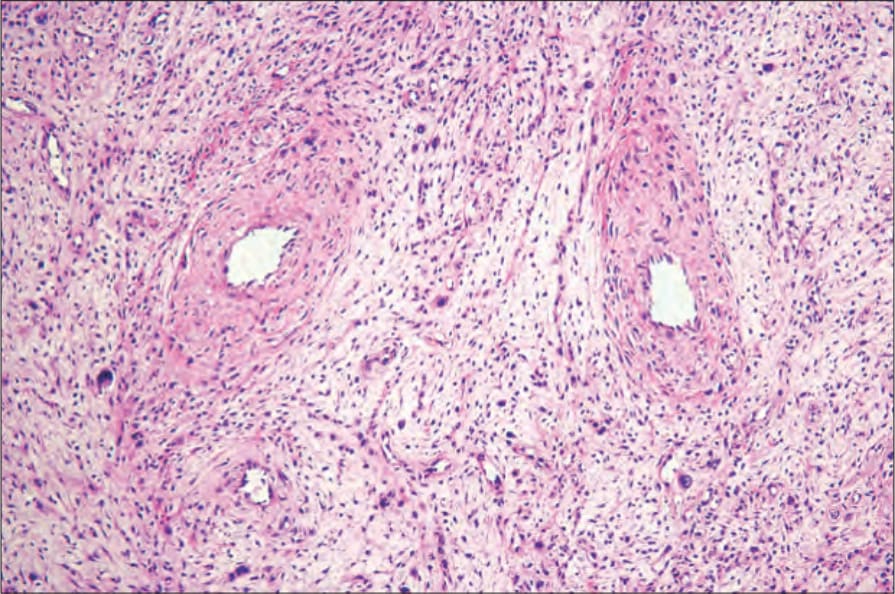
圖 12-283:纖維上皮性間質息肉 (fibroepithelial stromal polyp):可見厚壁血管 (thick-walled vessels) 伴隨細胞數量不一的疏鬆結締組織間質 (variably cellular loose connective tissue stroma)。By courtesy of M. Nucci, MD, Brigham and Women’s Hospital and Harvard Medical School, Boston, USA.
Fig. 12.283 Fibroepithelial stromal polyp: there are thick-walled vessels associated with a variably cellular loose connective tissue stroma. By courtesy of M. Nucci, MD, Brigham and Women’s Hospital and Harvard Medical School, Boston, USA.