Juvenile xanthogranuloma
Juvenile xanthogranuloma
This lesion usually affects the head and trunk but can also involve the genitalia (Fig. 12.281).1 Multifocal penile presentation has been documented,2 as well as a solitary perineal papule3 and a scrotal swelling.4 Clinicopathologically identical solitary lesions may be seen in adults.5 The histology is of lipid-laden histiocytes and giant cells, negative for CD1 and langerin, which are found in Langerhans cell histiocytosis. S100 may be positive in about 25% of cases. Juvenile xanthogranuloma is not associated with abnormal lipids but there may be a relationship with urticaria pigmentosa, diabetes mellitus, neurofibromatosis, cytomegalovirus infection, and leukemia.
A case of an isolated clitoral lesion is described.6
552 Diseases of the anogenital skin
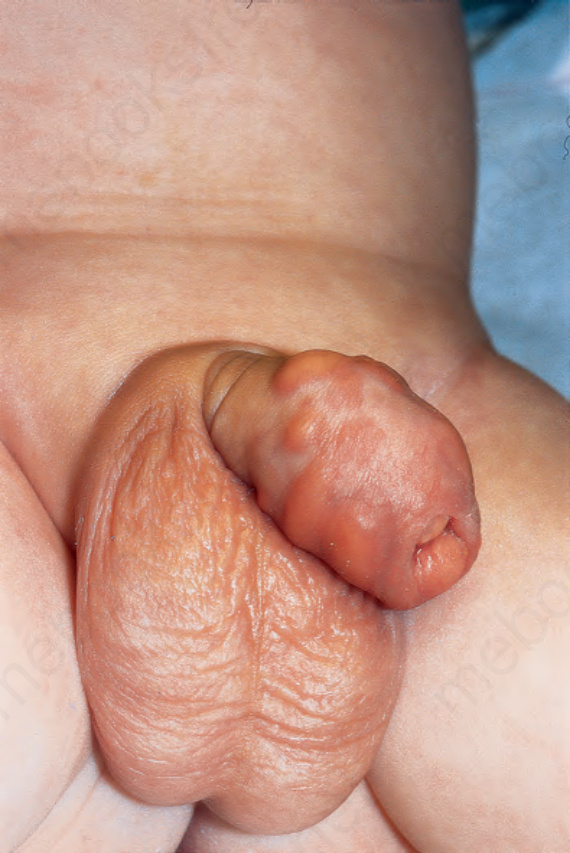
Fig. 12.281 Juvenile xanthogranuloma: lesions are present on the lateral shaft of the penis. Courtesy of R. Haufmann, Ulm, Germany. Reproduced with permission from Haufmann R.E., Bachor, R. Juvenile xanthogranuloma of the penis. J Urol. 1993:150:456–457. From Bunker C. Male Genital Skin Disease. Saunders Ltd./Elsevier 2004.
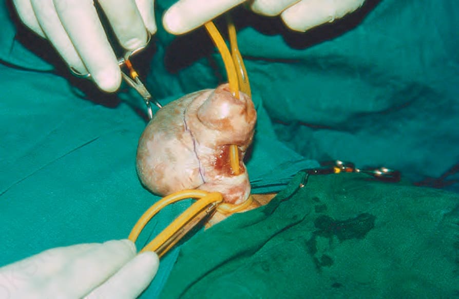
Fig. 12.282 Chronic edema simulating a keloid: note the dorsal proximal swelling and ventral urethral fistula. Courtesy of Dr Rameshwar Bang, Safat, Kuwait. Reproduced from Bang R.L. Penile edema induced by continuous condom catheter use and mimicking keloid scar. Scand J Urol Nephrol 1994;28:333–5. From Bunker C. Male Genital Skin Disease. Saunders Ltd./Elsevier 2004.
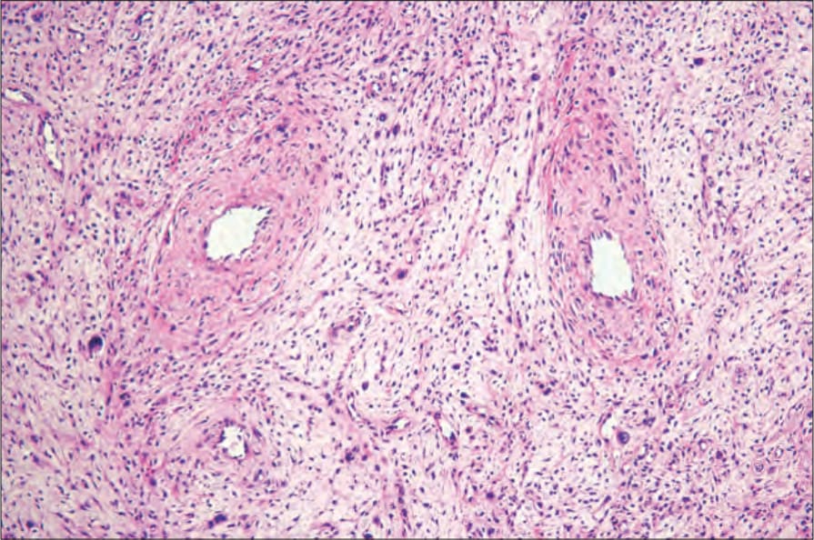
Fig. 12.283 Fibroepithelial stromal polyp: there are thick-walled vessels associated with a variably cellular loose connective tissue stroma. By courtesy of M. Nucci, MD, Brigham and Women’s Hospital and Harvard Medical School, Boston, USA.