Mouse papillomaviruses
Mouse papillomaviruses
While interest in human and nonhuman mammalian papillomaviruses has been waning since the commercialization of polyvalent recombinant vaccines, discovery of a mouse papillomavirus has opened up interesting new avenues for research. Numerous rodent papillomaviruses have been found in the past 30 years including one in a wild colony of European harvest mice.7–10 However, only recently has a laboratory mouse papillomavirus been reported in immunodeficient nude mice that could be transmitted to other nude mice but only to a limited number of inbred backgrounds. It was noted that pan T-cell deficiencies in the mice were needed for these lesions to develop. A very similar viral subtype was found in the normal skin of immunocompetent mice, suggesting that this virus has adapted well to its host. In the immunodeficient strains, benign papillomas (warts) developed on the muzzle and tail skin, but invasive, poorly differentiated carcinomas resembling trichomatrixomas developed on the dorsal lumbar skin (Fig. 36.8).11–15
In the mouse hair follicle, pigmentation follows a precise sequence of interactions between melanocytes and the dermal papilla.4,5 Hair is actively pigmented only during the anagen stage of the hair cycle. Melanin synthesis is inactive during catagen and remains so through telogen4; thus, it is important to emphasize that follicular melanogenesis is cyclic in nature.5
Currently, research in the mouse has identified more genes influencing coat color than any other trait.6 A search of Mouse Genome Informatics (www.informatics.jax.org) for the phrase ‘abnormal hair/coat pigmentation’ results in 1692 genotypes affecting coat color (http:// www.informatics.jax.org; August 2016).7 The most common alleles found in inbred research strains popular in research today are illustrated in Fig. 36.9. Although coat color genetics have been studied for nearly 100 years, the great variety of alleles affecting coat color appears to be a never-ending story. Current work in this field is actively pursuing the interconnectedness of the genes to form a genetic pathway (Fig. 36.10).
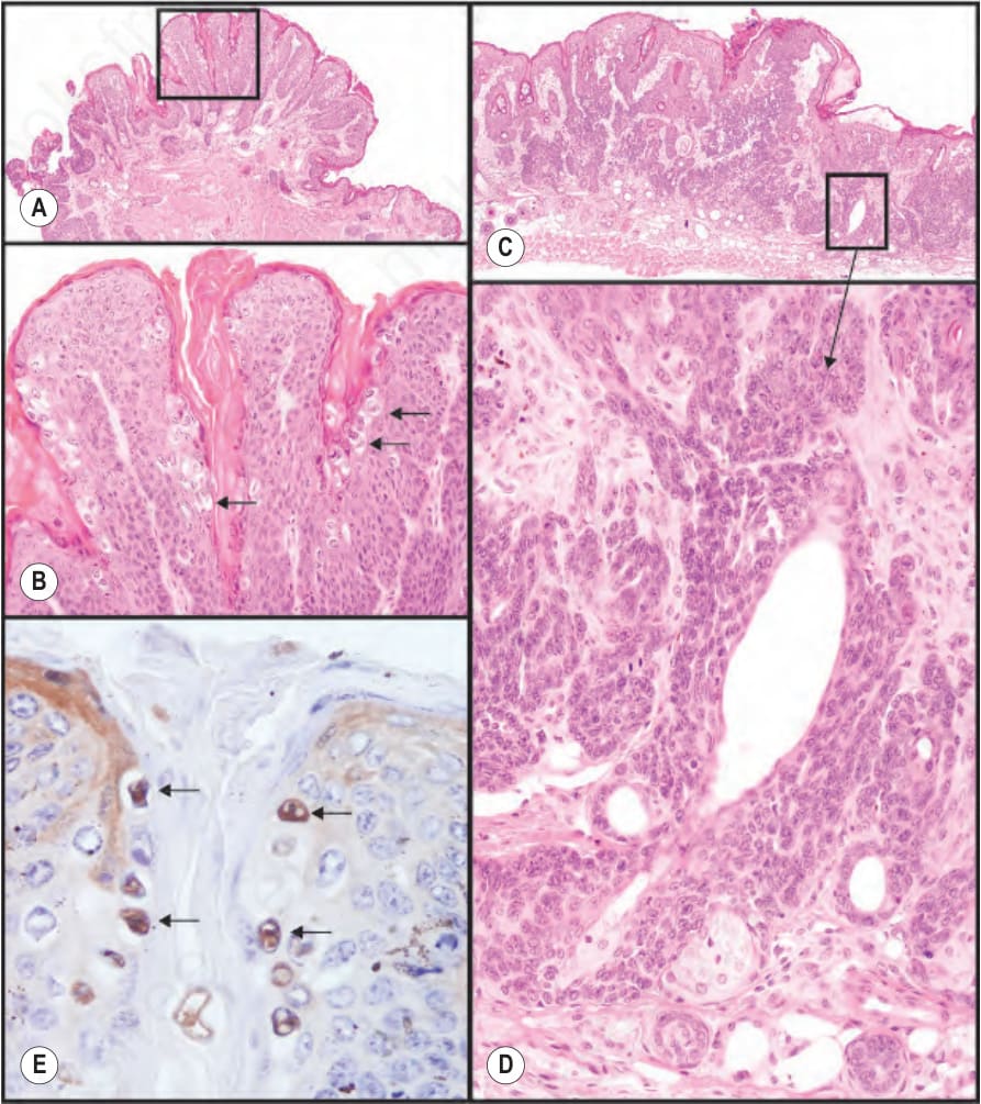
Fig. 36.8 Laboratory mouse papillomavirus infections cause exophytic papillomas on the muzzle (A, B) and tail that are productive (viral antigens can be detected in koilocytes by immunohistochemistry, E). However, when the virus was inoculated into the dorsal skin in the lumbar region, a slightly raised lesion developed with poorly differentiated cells that were locally invasive (C, D). These cells had a high nuclear to cytoplasmic ratio, high mitotic index, and were positive for several mouse specific keratins by immunohistochemistry.
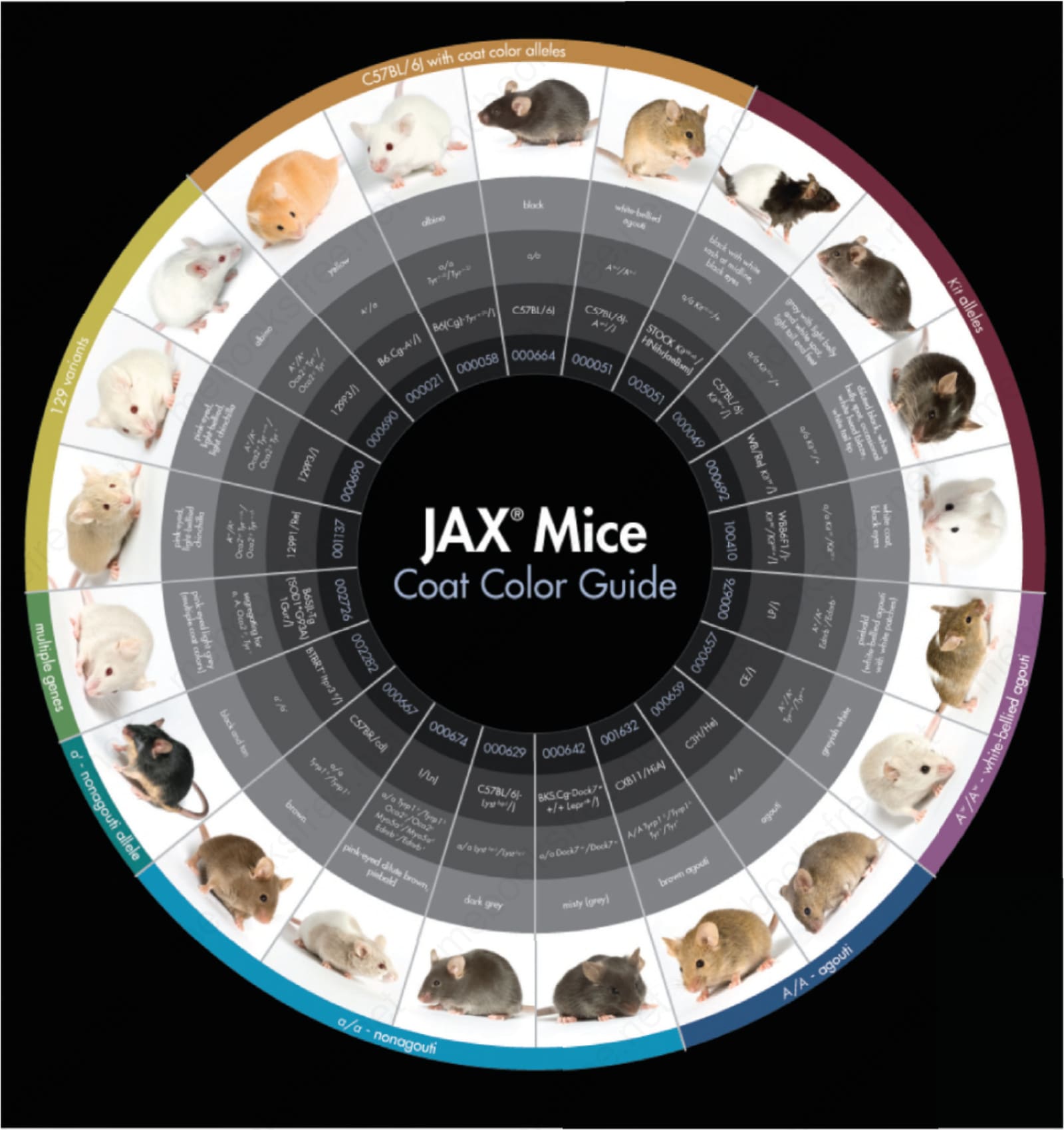
Fig. 36.9 Coat color variations in laboratory mice and the corresponding genetic mutations underlying the different colors. Stanton Short and Jennifer Torrance (JAX Creative Department, The Jackson Laboratory) are thanked for the figures and layout.
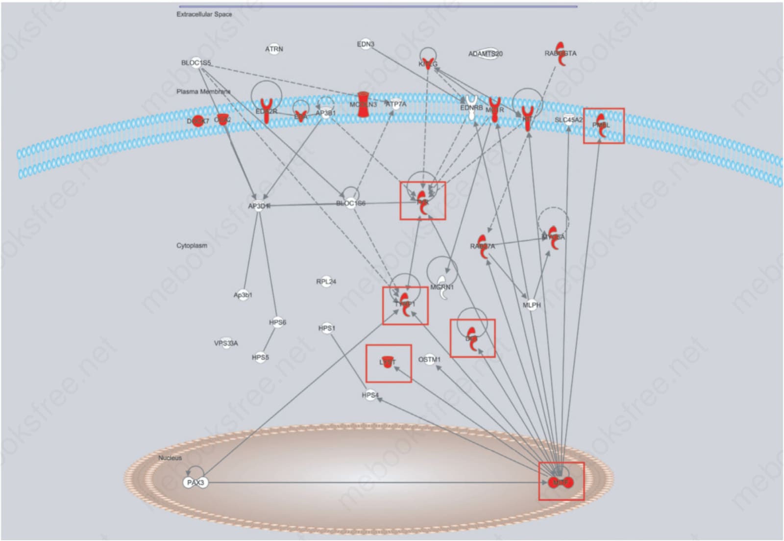
Fig. 36.10 Ingenuity Pathway Analysis Software® (http://www.ingenuity.com/) molecular pathway diagram for genes that interact in the coat color pathway. Many of the mutant mice in Fig. 36.9 have mutations in the genes in this pathway (red boxes). This illustrates how variations on a common trait, coat color, help to define a molecular pathway. It also illustrates the number of other genes involved with coat color not in this pathway, which will be future goals to integrate.