Hobnail hemangioma
Hobnail hemangioma
Clinical features Hobnail hemangioma (targetoid hemosiderotic hemangioma) usually presents on the limbs (particularly the thigh) and trunk of young or middle-aged adults and shows a male predilection.1–4 Lesions in infants and children are exceptional. Occasional tumors occur in the oral cavity including the tongue and gingivae.5,6 The lesion is asymptomatic, usually less than 2 cm in diameter, and increases in size very slowly. Patients sometimes describe cyclic changes.7 In women, lesions often become larger and darker prior to menstruation and become lighter and smaller after the menstrual period.8–11 In pregnancy, they increase in size probably due to estrogen, and in one case two lesions developed at the same time as the secondary sexual changes.9 Multiple lesions are exceptional. The original clinical description comprised a central red papule or macule, surrounded by successive clear and ecchymotic haloes (Fig. 35.491). Most often, however, the clinical presentation is nondistinctive and the differential diagnosis includes hemangioma, nevus or fibrous histiocytoma. There appears to be little or no tendency for recurrence.
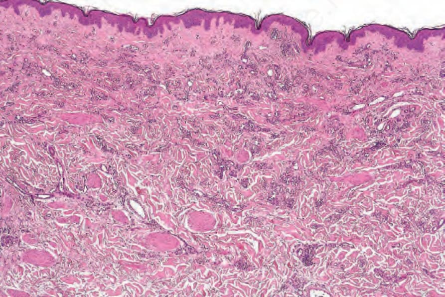
Fig. 35.489 Microvenular hemangioma: the manner in which the vessels irregularly infiltrate the dermis is sometimes mistaken for Kaposi sarcoma.
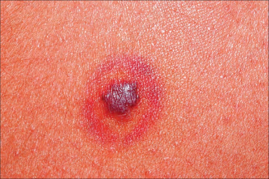
Fig. 35.491 Hobnail hemangioma: this example shows the characteristic targetoid appearance.