Piezogenic pedal papules
Piezogenic pedal papules
Clinical features Piezogenic pedal papules characteristically present as multiple skin-colored papules on the heels (Fig. 35.14).1–3 They show predilection for the internal aspect of the heels and are usually asymptomatic although pain may be elicited when the patient is standing. Lesions become more noticeable when the patient stands up as a result of pressure. Piezogenic pedal papules are common in patients with Ehlers-Danlos syndrome.4,5 They have been described in athletes, including a marathon runner, ice-hockey players, skaters and in association with Prader-Willi syndrome.6–9 Rare familial cases have been described.10 An association with mitral valve prolapse and mitral valve insufficiency has been reported.11–13
Histologic features The lesion is characterized by proliferation of mature fatty and fibrous tissue within the epineurium of a major nerve accompanied by prominent concentric perineural fibrosis (Fig. 35.15). In a handful of cases involving cervical and thoracic spinal nerves, the pathology was restricted to circumferential growth of fat around the epineurium.11 Rarely, bone formation has been described.12
Pathogenesis and histologic features It is likely that trauma plays a role in the pathogenesis of the lesions.6,7 A biopsy from a papule shows normal adipose tissue herniating into the dermis. This may mimic an intradermal lipoma but the clinical setting allows a diagnosis to be made. Rarely, prominent myxoid change and vascular proliferation can be seen.14
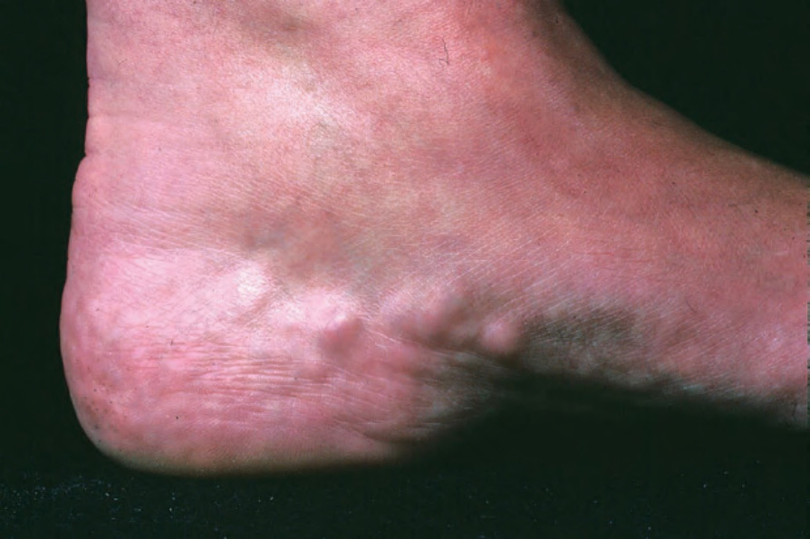
Fig. 35.14 Piezogenic pedal papules: note the multiple flesh-colored papules on the heel and lateral border of the foot. From the collection of the late N.P. Smith, MD, The Institute of Dermatology, London, UK.
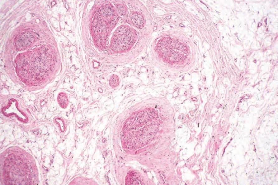
Fig. 35.15 Lipomatosis of nerve: note the extensive epineural fat deposition.