Pseudocyst of the auricle
Pseudocyst of the auricle
Clinical features Pseudocyst of the auricle (endochondral pseudocyst) is uncommon and shows a predilection for males.1–6 It presents as an asymptomatic, unilateral 1- to 5-cm swelling of the pinna.5 The scaphoid or triangular fossa of the antihelix is predominantly affected.7 Very occasionally, bilateral involvement may occur, and exceptionally the disease involves children.8–10 Although there is a predilection for the Chinese, other Asian races and Caucasians can be affected.11,12 If untreated, it may result in severe deformity of the ear.
Pathogenesis and histologic features The etiology is unknown. Low-grade trauma, ischemia, embryological defect of cartilage development, and autoimmunity have been suggested as possible causes.6,13 Repeated trauma in patients with ataxia has been associated with the condition.14 A recurrent case has been described in the setting of atopic dermatitis.15
Grossly, the pseudocyst may contain 1–2 cc of serous fluid, rich in lactate dehydrogenase-4 and -5.3,16 It is suggested that the presence of high lactate dehydrogenase levels results from trauma to the cartilage.6 Histologically, the lesion presents as an intracartilaginous cystic space lacking an epithelial lining (Fig. 34.46). Degeneration of the adjacent cartilage is often evident. There are no significant inflammatory changes.
Access ExpertConsult.com for the complete list of references
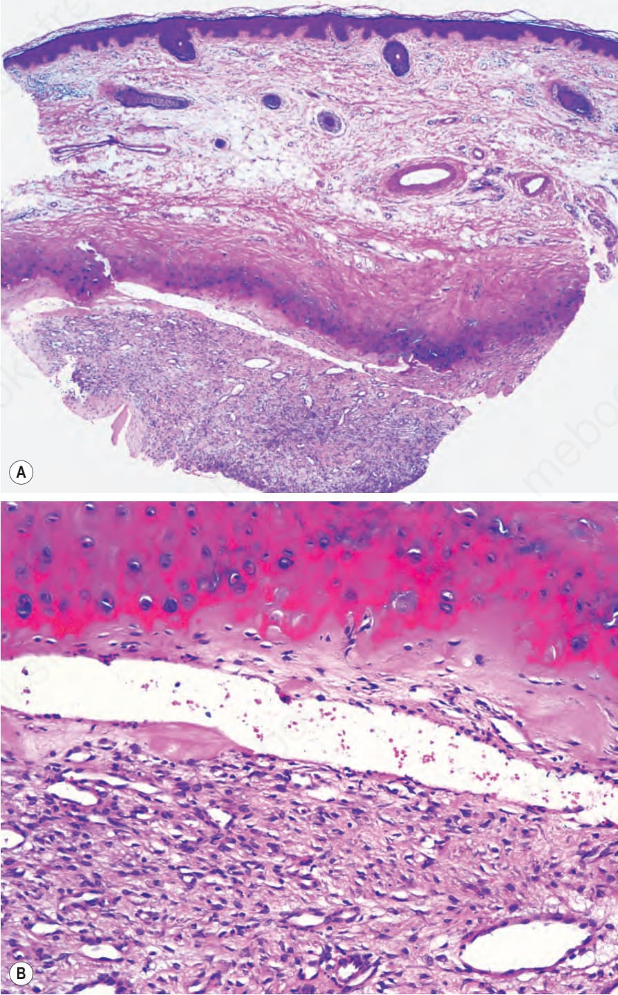
Fig. 34.46 Pseudocyst of the auricle: (A) cystic space within the cartilage; (B) the cavity is occupied by granulation tissue.